If you suffer from a very rare disease, getting the proper diagnosis can be an arduous journey. But a bigger challenge may be the feeling of isolation, since there may not be any support groups where you can connect to someone who is going through the same thing.
That was the situation the Bigelow family found themselves, and they turned to social media for the solution.
Bo Bigelow knew that his six-year-old daughter Tess had a genetic mutation called USP7. She also had global developmental delays in basic functions such as walking and talking, causing her to function at the level of an 18-month year old. Was USP7 the cause of her developmental delays?
Bigelow spread the word about his daughter’s genetic condition to find out, posting on Facebook, Twitter and a personal website with the plea to “help us find others like Tess.” A friend of the family also posted on Reddit, where it was read within 24 hours by a researcher at Baylor College of Medicine who was studying USP7. His research group had already identified seven children similarly affected by the same genetic mutation, and they were about to publish an article about it in Molecular Cell.
Tess may become part of future clinical trials at Baylor, but the researchers also connected the Bigelows to the other seven families. “These days there are ribbons and awareness-weeks for so many diseases,” Bigelow said in a recent KQED Science story, “but when yours is ultra-rare, you feel completed isolated. You feel like you’re never going to hear another person say, ‘Us too!’ And being connected to other families changes all that.”
The KQED piece goes on to explain:
“Patients or parents like Tess’ who are seeking answers to seemingly unsolvable medical mysteries have new tools to reach out, not only on social media, but in crowdsourcing websites like CrowdMed, a subscription service for people seeking answers to medical conundrums. At CrowdMed, people who have symptoms but have yet to find a diagnosis seek opinions from the site’s “medical detectives,” only some of whom are medical professionals.”
This is a reposting of my Scope blog story, courtesy of Stanford School of Medicine.
Space is a hostile place, even inside a spacecraft. Radiation, weightlessness and isolation are only a few of the unique stressors faced by astronauts during space travel.
As NASA prepares for a manned journey to Mars, researchers are studying what happens to the human body in space to determine the health risks of a several-year mission. This research includes a unique study of identical twin astronauts to investigate the effects of spaceflight at a molecular level — comparing data from Scott Kelly, who recently completed a one-year space mission, with data from his brother who led a normal life on Earth.
NASA recently produced a series of web videos, “Omics: Exploring Space Through You, ” that discusses its twins study and features Michael Snyder, MD, professor and chair of genetics at Stanford and principal investigator on one of the projects. Omics is a field of study that integrates different types of molecular information and, as Snyder explains in the introductory video:
“In many respects, it’s like a jigsaw puzzle. A jigsaw puzzle can be made of 1000 pieces but you don’t really see the picture until you put all those pieces together. That’s the same for omics; you basically try and understand all of the individual pieces so you can see the whole picture.”
NASA is making billions of measurements of both twins to see what space really does to the human body. And researchers hope that one day omics profiles will be conducted on a large scale in clinics, not just on astronauts, so we can switch from a “one size fits all” approach to personalized medicine.
“OMICS is really an amazing field where we can look at people and their health at a level that’s never been possible before,” Snyder comments. “And with that we’ll be able to better manage people’s health and try and keep them healthy long before they get sick.”
This is a reposting of my Scope blog story, courtesy of Stanford School of Medicine.
How do you combine internal medicine, nutrition, culinary arts and public policy into a single career? Ask Michelle Hauser, MD, who has integrated her eclectic training into a unified research program to help improve the health and wellness of people in underserved communities.
Although Hauser always dreamed of being a physician, she began her career as a Le Cordon Bleu chef with a culinary internship at Alice Water’s famous restaurant, Chez Panisse. Hauser then put herself through college at Humboldt State University by teaching at local cooking schools, before going on to Harvard to earn her MD and MPA.
Hauser is now a postdoctoral research fellow in cardiovascular disease prevention at the Stanford Prevention Research Center and practices primary care at Fair Oaks Health Center, a clinic for those with limited access to health care in Redwood City. She is also on the board of directors of the American College of Lifestyle Medicine. I recently spoke with Hauser about lifestyle medicine and medical care for the underserved:
Why did you become a chef when you dreamed of being a doctor?
I grew up poor in rural Iowa, without a support system or parents who had gone to college. My high school guidance counselor actually laughed at me when I told her I wanted to be a doctor. She said, “let’s find something more suitable for you to do,” and went on to suggest that I work at a local factory.
So I went to culinary school because I love to cook. And everyone told me that I’d never be a doctor, so I thought maybe they were right. As I finished culinary school, however, I knew that I wasn’t afraid to fail at my dream of becoming a physician. I would, however, regret not trying.
How did you become interested in treating medically underserved patients?
Coming from an underserved background inspired me to focus on medical care for the underserved. Additionally, I became very interested in the prevention of chronic disease —particularly via lifestyle changes — and the disparities in access to preventive care and services.
I’m currently involved with Fair Oaks Health Center’s care transformation project to increase and improve wellness resources for patients with metabolic risk factors for cardiovascular disease and diabetes. All resources and classes are available in Spanish and English. It’s truly rewarding to work with a diverse group of physicians, nurses, dieticians, psychologists and health educators to brainstorm and test new models of chronic disease prevention and treatment in this type of underserved clinical setting.
What is lifestyle medicine?
Lifestyle medicine is a field of medicine that encompasses research, prevention and treatment of disease caused by lifestyle factors such as nutrition, physical inactivity, smoking, excessive alcohol use, poor sleep and chronic stress. These lifestyle factors are currently responsible for nearly 80 percent of both chronic diseases and healthcare spending.
A goal of the American College of Lifestyle Medicine, and lifestyle medicine in general, is to improve personal and population health — adding both life to years and years to life. I was inspired to join the board of directors of ACLM because I fervently believe that healthcare needs to better address the root causes of disease and not just treat the downstream effects.
How can you motivate people to make lifestyle changes and stick with them?
This question is the elephant in the room. While there are many examples of people — myself included — who have turned their lives around with improved lifestyle habits, we have yet to find the perfect set of instructions for everyone to change their behaviors.
However, there are many promising techniques out there and much research currently being done. For instance, Brian Wansink, PhD, professor and director of the Cornell University Food and Brand Lab, has done a lot of research investigating how we constantly make mindless choices — particularly about what and how much to eat. He has shown that if we swap healthy items for unhealthy ones in the places that we’re most likely to select our food, we won’t even notice that we’ve suddenly opted to eat healthier.
We also need to change the way preventive care and lifestyle-based treatments for chronic diseases are paid for. Unless government and private insurance programs reimburse these services, most people will not have access to them.
How do you use nutrition and culinary education in your practice?
When I was in medical school, we piloted and evaluated a program that used shared medical appointments that incorporated cooking demonstrations and nutrition classes with primary care management for patients with cardiovascular risk factors. We found the program to be feasible, cost-effective and well received by patients.
I went on to conduct similar group sessions in my residency primary care clinic, and am now working on several projects that utilize cooking skills for disease prevention and treatment in my current practice and research.
With your busy work life, do you still have time to cook for yourself?
Absolutely! I cook most of the meals that I eat. These are not generally the fancy fare that I prepared in restaurants or culinary classes, but satisfying, delicious and healthy, all the same. I occasionally post about these recipes and answer food and nutrition-related questions on my blog, Chef In Residency. Pictures of other quick meals that I make on weekdays can be found on my Chef In Residency Instagram page.
This is a reposting of my Scope blog story, courtesy of Stanford School of Medicine.
Poor sleep is likely to make you feel grumpy and unfocused, but more importantly it puts you at risk for serious medical conditions such as obesity, type 2 diabetes and heart disease — and it shortens your lifespan.
A new study shows that sleep loss also increases your risk of cardiovascular disease by changing how your body metabolizes cholesterol.
Recently reported in Scientific Reports, University of Helsinki’s sleep team studied the cholesterol metabolism of sleep-deprived people, measuring their gene expression and the levels of blood lipoprotein, a molecule that transports cholesterol through the blood. They assessed these factors for a small group of volunteers who only slept four hours per night for five days. The team also looked at longer-term effects on cholesterol metabolism using data from two large population studies with 2739 participants.
The study found that people getting insufficient sleep have fewer high-density lipoproteins (HDL) — the “good” proteins that act as cholesterol scavengers to decrease accumulation of atherosclerosis within the walls of arteries — than people who get enough sleep.
“It is particularly interesting that these factors contributing to the onset of atherosclerosis, that is to say, inflammatory reactions and changes to cholesterol metabolism, were found in the experimental study and in the epidemiological data,” said Vilmo Aho, a graduate student at the University of Helsinki, in a recent news release.
The bad news is they showed that even a week of sleep deprivation had a significant impact. Aho explained in the release:
The experimental study proved that just one week of insufficient sleep begins to change the body’s immune response and metabolism. Our next goal is to determine how minor the sleep deficiency can be while still causing such changes.
This is a reposting of my Scope blog story, courtesy of Stanford School of Medicine.
New research shows that familial hypercholesterolemia — a genetic condition that leads to high LDL cholesterol — is commonly diagnosed late and patients often don’t get adequate treatment. FH can cause aggressive and premature heart disease, including heart attacks, strokes, narrowing heart valves and sudden cardiac death.
Joshua Knowles, MD, PhD, assistant professor of cardiovascular medicine and chief medical advisor for the FH Foundation, is senior author of a new study that characterizes adult FH patients in the United States using data from the new CASCADE FH Registry™. As reported in Circulation: Cardiovascular Genetics, the study found many flaws in the current treatment of patients with FH. I spoke with Knowles about this silent and deadly disease:
What is Familial Hypercholesterolemia?
Familial Hypercholesterolemia is the medical term given to very high cholesterol that runs in families. We say, ‘We never find an individual with FH. We only find families with FH.’
It’s caused by genetic mutations that control the body’s ability to recycle LDL cholesterol. Your liver makes cholesterol and sends it through the bloodstream. Your body takes what it needs and then sends excess LDL cholesterol back to the liver for recycling.
In FH patients, the liver cannot recycle LDL cholesterol because there are defects in some receptors that pull the cholesterol from the blood. Therefore, the LDL levels remains very high in the blood, which is toxic to the blood vessels over time. If you have FH, your LDL cholesterol levels are two to three times higher than normal and that puts you at a much, much higher risk of early onset coronary disease such as heart attacks.
How is FH diagnosed?
There are over a million people in the US with this disorder, but less than ten percent have been diagnosed. In the US, genetic testing hasn’t become standard of care yet, largely due to the cost. So usually FH is diagnosed with a clinical point system based primarily on your personal and family medical history.
If you’re not diagnosed and treated, your risk of a heart attack is extremely high. However, if you are diagnosed, you can be treated and live a long and healthy life. So it’s a poster child for preventative and personalized medicine.
How is FH treated?
Lifestyle changes — like improving your diet or exercising — are almost never enough for FH patients. FH patients need to be treated with medications that lower their LDL cholesterol. For most people with high cholesterol, one drug is sufficient. However, many FH patients require more than one drug.
The most common and important medications for FH are statins, which work by tricking the body into activating the LDL recycling program. Normally for every gene, you have one copy from Mom and one copy from Dad. In the most common form of FH, you’ve inherited the mutation from one parent. So you have one receptor in the liver that works well and one that doesn’t work at all. The statin drug tricks the body into increasing the levels of the good receptors to compensate for the bad ones — basically putting the good receptors in overdrive to activate the LDL recycling program.
What do you recommend for patients with a family history of early heart disease?
Really the biggest and best thing is to get your cholesterol tested when you’re young, when nothing but the genetic condition causes high cholesterol. If you wait until your 60 years old, it can get trickier to figure out and may be too late. Guidelines from the American Academy of Pediatrics recommend cholesterol screening for children as young as ten years old for the general population and as young as two years old for families with a history of FH. Current research shows that intervening early has a big impact.
What is the CASCADE FH Registry and why is it important?
To advocate for change, it’s really important to have some numbers to back up what we’re saying. When we publish on the data, it raises the profile of the condition. We also need a baseline to compare to in the future. The beauty of the new CASCADE FH Registry is that we’ll be able to follow patients over time to see if what we are doing is making a difference.
It’s not just important for the individual to know whether they have FH. It’s also important for the family to know. When you identify one person with FH, the real potential benefit comes with screening the rest of the family so you can identify ticking time bombs before they have problems. This is called cascade screening – hence the name of the registry. We want to prevent people with FH from becoming patients with FH. We want to prevent those heart attacks.
This is a reposting of my Scope blog story, courtesy of Stanford School of Medicine.
Despite careful patient selection, only about 75 percent of heart recipients survive three years after the transplant surgery. Identifying the patients most in need of additional care has always been tricky, but now Stanford researchers have found a better way to predict which heart transplant recipients have a higher risk of dying or needing another heart transplant, as reported in Circulation today.
One key reason transplant patients die is cardiac allograft vasculopathy, an accelerated and aggressive form of coronary artery disease.
William Fearon, MD, professor of cardiovascular medicine and senior author, explained the significance of their results in a recent email:
Identifying patients at higher risk of dying from cardiac allograft vasculopathy is helpful, because it allows the transplant physicians to be more aggressive with medical therapy and monitoring than they might otherwise be, in order to hopefully prevent adverse events.
The researchers conducted a clinical trial involving seventy-four heart transplant recipients, whose heart physiology was invasively assessed within eight weeks and one year after transplantation. They found that two particular diagnostic procedures were able to successfully identify high-risk recipients — fractional flow reserve and index of microcirculatory resistance.
Fractional flow reserve is a procedure that measures the blood pressure and flow through a specific part of the coronary artery. It is often used to determine whether blood flow is significantly obstructed by a blockage or lesion, guiding a cardiologist’s decision of whether to stent the blockage.
Fearon’s team determined that a low fractional flow reserve measured soon after the transplant independently predicted the heart transplant recipients’ risk of death or retransplantation.
Index of microcirculatory resistance measures the functionality of the tiny vessels that supply blood to the heart, such as capillaries, arterioles and venules. Fearon found that a higher than normal reading measured one year after the heart transplant was also an independent predictor of the recipients’ event-free survival.
The Stanford researchers hope that more emphasis will be placed on these two invasive assessments of cardiac physiology in heart transplant recipients, so their medical regimen can be adjusted to improve the odds of their survival.
This is a reposting of my Scope blog story, courtesy of Stanford School of Medicine.
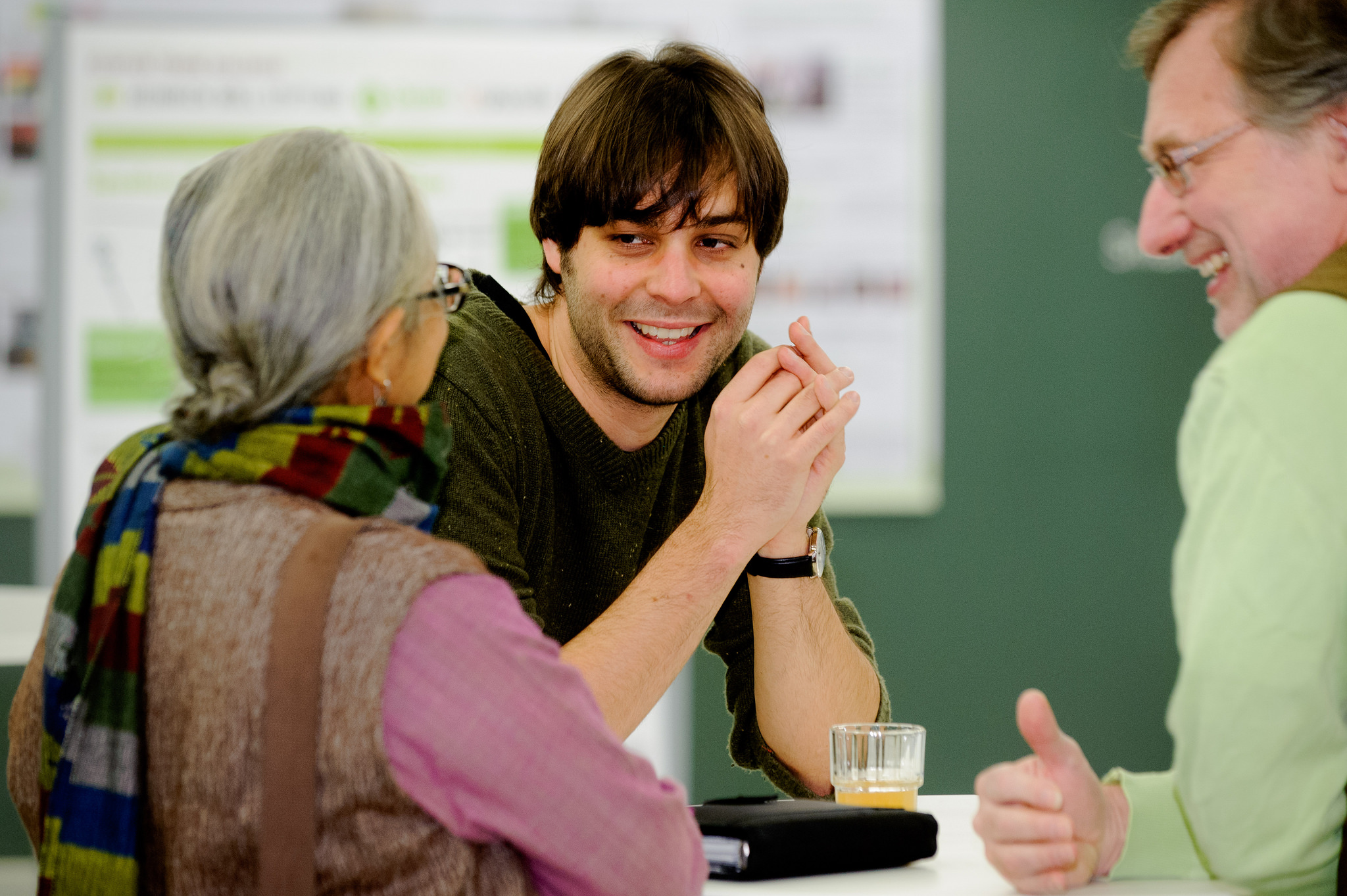
Just about everyone I know feels overwhelmed with an endless ‘to do’ list of work assignments, chores, errands and appointments. By the end of the day, we often don’t feel up to hitting the gym or going out. We just want to go home to collapse and recharge.
Our busy lives can make it hard to spend much quality time with friends and family. However, research suggests that we need to make this a priority, because social connections impact our health and wellness.
Many studies show that there are distinct, positive physical and emotional benefits from having supportive social connections. Research suggests that illness rates are lower, as is premature death, for those who are socially connected. What seems eminently clear is that positive, supportive connections help people manage the stress of daily living better than people who do not have the outlet of someone who will listen and empathize with their experience.
Gomperts recommends several ways to expand your social connections, such as joining a book club or knitting club, taking a class or volunteering. The key is to find something that works for you. She explained:
For some people, getting out can be really hard — whether due to depression, social anxiety or a lack of time due to pressures in life. However, finding a way to connect is incredibly important, and there are Internet options that can be very useful.
Some people can also benefit from sharing their sense of isolation, loneliness or desire to have more social connections with a counselor or support group — such as those offered at the Faculty Staff Help Center.
This is a reposting of my Scope blog story, courtesy of Stanford School of Medicine.
One of the oldest scientific debates is “nature versus nurture” — do inherited traits or environmental factors shape who we are, and what we do?
So far it’s a draw.
For instance, a massive meta-study, reported in Nature Genetics, quantified the heritability of human traits by analyzing more than 50 years of data on almost 18 thousand traits measured in over 14.5 million pairs of twins. They determined that heritability accounted for 49 percent of all traits and environmental influences for 51 percent.
They essentially found that genes and the environment play an equal role in human development. But that isn’t the end of the debate.
The researchers used PET imaging to measure the glucose — or energy — metabolism throughout the brain. The authors explained their motivation in the JNM article:
“The patterns of glucose metabolism in the brain appear to be influenced by various factors, including genetic and environmental factors. However, the magnitude and proportion of these influences remain unknown.”
The researchers studied 40 identical twin pairs and 18 fraternal twin pairs. Any differences between identical twins is expected to be due to environmental factors since they are genetically identical, whereas fraternal twins only share half the same genes on average.
The researchers compared imaging results between the two types of twins to estimate the extent of genetic and environmental influences. When a genetic influence is dominant, the identical twins would have more trait similarity than fraternal twins. When an environmental influence is dominant, the trait similarity would be the same for identical and fraternal twins.
The researchers found that both genetic and environmental factors influenced glucose metabolism in the brain, but they predominantly affected different areas. Genetic influences played a major role in the left and right parietal lobes and the left temporal lobe, whereas environmental influences were dominant in other regions of the brain.
The brain’s parietal lobes process sensory information such as taste, temperature and touch, and the temporal lobes process sounds and speech comprehension. More research is needed to understand why these areas of the brain where influenced more by genetics.
In addition to adding new information to the “nature verses nurture” debate, these results could be applied to other research areas, such as using imaging to better understand the underlying cause of Alzheimer’s disease or psychiatric disorders. Identifying which regions of the brain are more influenced by genetics or the environment may add critical information to help better understand and treat diseases.
This is a reposting of my Scope blog story, courtesy of Stanford School of Medicine.
Stanford chemists have now developed a highly sensitive and specific tool to screen cancer and HIV — 1000 times more sensitive than current clinical tests. More precise screening could allow for much earlier detection and treatment, as well as help avoid false positive results and their resulting unnecessary procedures and stress.
Developed in the lab of chemist Carolyn Bertozzi, PhD, this new ultrasensitive screening technique has already been tested as a biomarker for thyroid cancer in clinical trials. The study results were recently reported in ACS Central Science.
Many standard clinical blood tests are based on immunoassays, which use highly specific antibodies to detect specific molecules known to be associated with the target disease. Bertozzi’s new screening test adds the power of DNA detection to this standard procedure. Rather than marking, or “flagging”, disease-related antibodies using customary chemical compounds, the team flagged the antibodies using DNA.
The researchers tested their technique, with its signature DNA flag, against four commercially available, FDA-approved tests for a biomarker for thyroid cancer. It outperformed the sensitivity of all of them, by at least 800 times, and as much as 10,000 times. By detecting the biomarkers of disease at lower concentrations, physicians could theoretically catch diseases far earlier in their progression.
Bertozzi is currently testing their ultrasensitive screening method in clinical trials for other diseases, including HIV. If its effectiveness is proven, the researchers expect it to be readily adopted in clinics. Cheng-ting “Jason” Tsai, co-author and graduate student in Bertozzi’s group, said in the news release:
Many of our collaborators are excited that the test can be readily deployed in their lab. In contrast to many new diagnostic techniques, this test is performed on pre-existing machines that most clinical labs are already familiar with.
This is a reposting of my Scope blog story, courtesy of Stanford School of Medicine.
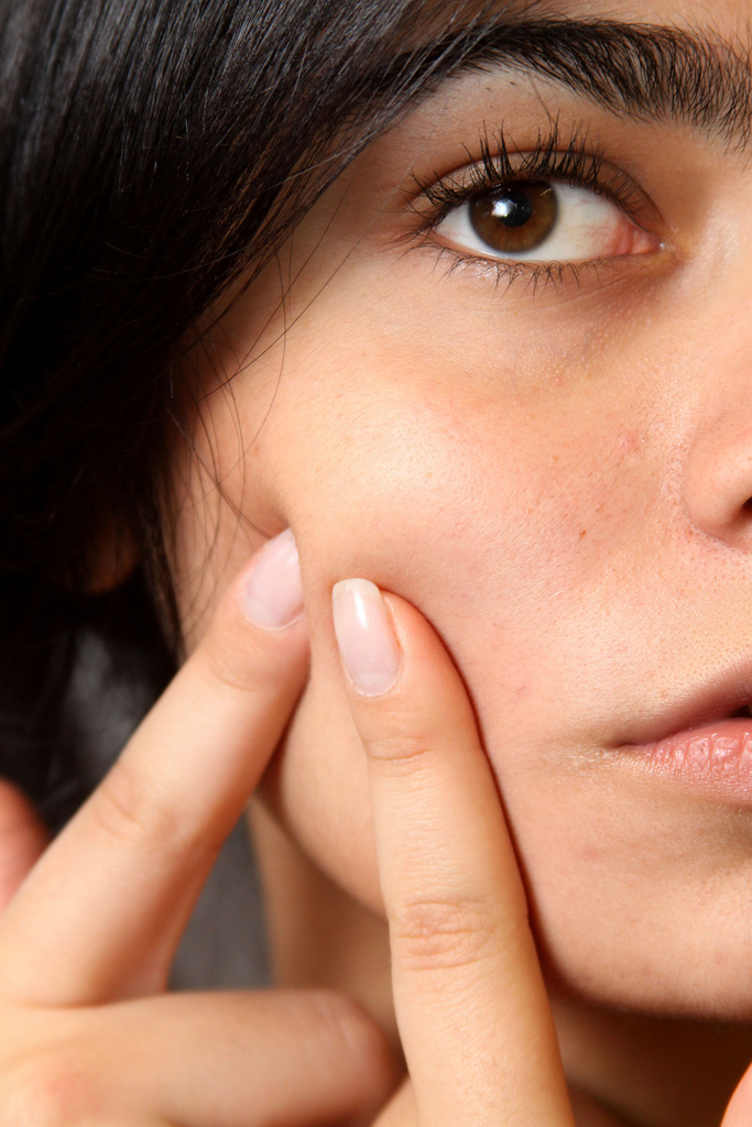
Most of us suffer through at least minor acne as a teenager, but many battle severe acne into adulthood. It affects up to 50 million people annually and can cause permanent scarring, poor self-image, depression and anxiety.
The American Academy of Dermatology recently published new guidelines for acne treatment in the Journal of the American Academy of Dermatology. The new guidelines recommend using several therapies at once. I spoke about them with Justin Ko, MD, MBA, clinical assistant professor of dermatology.
What is your advise for people that suffer from mild to severe acne?
There are great treatments out there! Find a physician with whom you feel comfortable; someone who is willing to talk through the reasons behind acne and formulates with you a personalized treatment approach based on your type of acne, skin type, other health issues, preferences, etc. A therapeutic partnership between a provider and patient is essential. I think that being able to treat acne successfully and effectively is the mark of a good dermatologist. It is and remains immensely satisfying for me to go through this journey with my patients and see them come out the other side with a newfound comfort in their skin.
What do you think of the new American Academy of Dermatology guidelines?
The AAD’s acne treatment recommendations represent the current standard of care. Our core treatment arsenal is comprised of topical treatments, oral antibiotics, hormonally-based treatments and isotrenoin (accutane), as well as other less-commonly used treatments that can have their place for the appropriate patent or situation. I agree with the guidelines that it is especially important for a topical regimen to form the foundation of any approach to acne treatment and not to rely on a single modality.
How has acne treatment changed in the past two decades?
We now have an appreciation for the fact that different types of acne require different approaches. I myself am using oral antibiotics less and more hormonally-based treatments and isotrenoin (accutane) when I think a patient will benefit. Here at Stanford, we also see acne-like eruptions in different forms due to underlying medical conditions and treatments, including new targeted-cancer treatments.
How do laser treatments and photodynamic light therapy work?
In the right settings these treatments can be good, as a companion to “traditional” treatments or situations when a patient is unable to use “traditional” treatments. They work in a couple ways. Some light-based treatments take advantage of a property of selected wavelengths of light that reduce the skin’s immune activity. Acne is fundamentally inflammation around hair follicles, so these light-based treatments can help.
Photodynamic therapy and intensive treatment protocols for PDT actually aim to shrink the oil glands, which play a role in acne formation in unlucky people. This intensive treatment can be quite painful, but it can be effective.
As with any of this, it’s essential to find a provider who is trained in the appropriate, safe and effective use of laser therapy.
This is a reposting of my Scope blog story, courtesy of Stanford School of Medicine.


