Stanford otolaryngologist champions ultrasound imaging

Photo, of Lisa Orloff performing an ultrasound exame, by Stuart Kraybill
Photo, of Lisa Orloff performing an ultrasound exam, by Stuart Kraybill

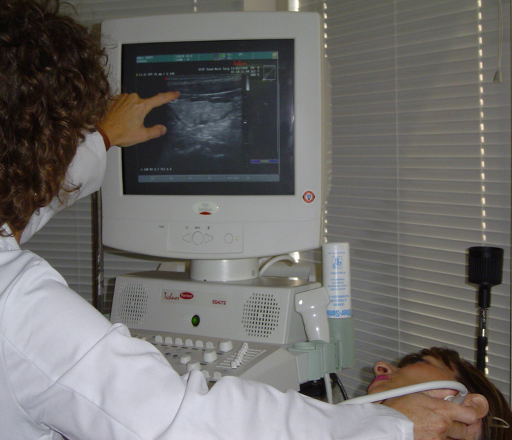
Patients with thyroid nodules — extremely common lumps on the thyroid that are usually benign, but can be malignant — are typically sent for ultrasound imaging to evaluate the size and structure of their thyroid and nodules. A radiologist’s report is then sent to the treating physician, who discusses the report with the patient and recommends next steps.

Lisa Orloff, MD, director of the endocrine head and neck surgery program at Stanford, doesn’t follow this traditional procedure: she performs her own ultrasound exams in the office and is training other head and neck surgeons to do the same. I recently spoke with Orloff about the role of ultrasound imaging in her practice.

Why do you primarily use ultrasound imaging to diagnose head and neck disease?

“My clinical practice focuses on the surgical management of thyroid and parathyroid disease, especially thyroid cancer. In the head and neck region, ultrasound imaging has long been recognized as the ‘go to’ study if you want to evaluate the structure, size and content of the thyroid gland. What’s been recognized more recently is how great ultrasound is for most of the head and neck structures. So we’re moving into an era of ‘ultrasound first:’ See what you can see with ultrasound, and then decide if you need additional cross-sectional imaging to corroborate or complement the ultrasound findings. For patients with thyroid cancer, ultrasound is extremely useful for evaluating not only the thyroid, but the rest of the neck for aggressive features including possible metastases.

Ultrasound is a low risk, low cost and very high yield imaging study that better characterizes the details within thyroid nodules or lymph nodes; whereas, CT and MRI often rely more on size to say whether or not a thyroid nodule or lymph node is suspicious. It’s really phenomenal what you can see with modern, high-resolution ultrasound equipment.

However, ultrasound has been blamed for the recent increase in incidence of thyroid cancer, which is largely due to increased detection. Even malignant thyroid nodules can sometimes be very indolent cancers that may not require intervention, but can be monitored. A major challenge in thyroid cancer care is distinguishing potentially aggressive tumors from those that are very low risk.”

Why is it helpful to have a clinical doctor, instead of a technician, perform ultrasound?

“When used at the point of care — performed by the clinician who is taking care of the patient — ultrasound enables the treating clinician to immediately investigate and answer questions with ultrasound information, and then implement treatments. It’s sort of one-stop shopping.

There’s an invaluable connection made with the patient when the treating physician performs the ultrasound exam, while explaining findings to the patient and discussing whether and how to treat them. I think it translates into improved patient care. If I’m the one doing the ultrasound exam, I can plan and execute surgery better with first-hand knowledge of what lies beneath the surface — rather than relying on images that someone else captured. I can perform ultrasound-guided biopsies and treatments in the office. I can also judge firsthand when an intervention or even biopsy isn’t necessary.

At present, I’m the surgeon in the head and neck division who routinely uses office-based ultrasound to evaluate patients, many of whom are referred to me specifically for that reason. But my colleagues in comprehensive ENT also perform ultrasonography [ultrasound imaging], as do our fellows and residents. We’re very motivated to train the next generation of otolaryngologists so it becomes more widely practiced in the office setting. We want to reduce the need for multiple appointments and more costly or invasive studies.”

I heard you recently traveled to Zimbabwe. What did you do there?

“My department has developed a relationship with the only medical school in the country, the University of Zimbabwe. I spent two weeks this summer, mainly teaching ultrasonography to residents in both otolaryngology and surgery — introducing the concept of point-of-care ultrasound to a low-resource practice environment where this has the potential for even greater impact. Most patients there don’t have ready access to get an expensive CT or MRI scan. I think ultrasound has a particular application in that setting, because it’s inexpensive, portable, fast and so user friendly. It’s also painless and non-threatening — you can do it on kids without having to anesthetize them to stay still.

Going over there to teach was a really rewarding experience. I hope to go back soon. We were very fortunate to have ultrasound equipment loaned for teaching purposes by GE based in South Africa. My next goal is to raise funds for an ultrasound machine to equip the Zimbabwe program with this wonderful tool for their continuing use.”

This is a reposting of my Scope blog story, courtesy of Stanford School of Medicine.

Chronic pain is correlated with major depression — for sufferer and spouse, study finds

Screen-Shot-2016-08-24-at-11.29.29-AM-e1472063483801-300x283
Image by Stefano Vitale via Stanford Medicine magazine

Chronic pain — pain that lasts longer than three months — debilitates over 100 million Americans and costs the United States over half a trillion dollars annually, but we still don’t really understand its underlying risk factors.

According to a new study recently published in PLOS Medicine, genetics and your home environment substantially contribute to your risk of chronic pain. The study also found that chronic pain is correlated with major depressive disorder (MDD), and both conditions are in part caused by a variety of genes.

The study was conducted by Andrew McIntosh, MD, chair of biological psychiatry at the University of Edinburgh, and his colleagues using data from two large United Kingdom population studies — including 23,960 individuals from the Generation Scotland: Scottish Family Health Study and 112,151 individuals with genotyping and phenotypic data from the United Kingdom Biobank.

It makes sense that someone with chronic pain has a higher risk of being severely depressed. But the study found that you also have increased risk for major depression if you’re living with a spouse or partner with chronic pain.

The authors discuss possible reasons for this spousal effect, which were summarized in a recent news story:

  • “You may choose a spouse similar to yourself, with similar existing predispositions to the conditions (assortative mating).
  • It’s possible that caring for a spouse with chronic illness makes you more likely to develop depression.
  • The environment you share with your spouse may contribute to both your risks of chronic pain and MDD; shared environmental factors could include diet, infectious disease, and hobbies.”

Determining the extent of these environmental factors was beyond the scope of the current study, but the authors recommend future research to identify the causal mechanisms that link chronic pain and major depressive disorder. They concluded in the paper:

“The answers to these questions are likely to signpost new directions for therapeutic interventions and highlight the symptoms that are most amenable to treatment, as well as prevention.”

This is a reposting of my Scope blog story, courtesy of Stanford School of Medicine.

Looking beyond life as a bioscience university professor

Photo by Vic
Photo by Vic

Life as a tenured university professor seems idyllic with its job security, intellectual freedom, prestige, livable wage and flexible schedule. No wonder so many bioscience students aim to become professors.

But numerous factors, including a lack of available faculty positions, are making bioscience trainees consider other careers. That’s been the experience for Scott Carlson, PhD, a Stanford postdoctoral research fellow in biology, who recently told me:

“My dream job is a baffling question right now. When I started as a postdoc, I would have said my dream job was to be a professor at a program in interdisciplinary biology or bioengineering. After five years as a postdoc, I’m not sure anymore but I don’t know what to replace it with. Academia makes it impossible to explore other options. If I leave, my grants would disappear and it would be hard to get back in without recent publications.”

Carlson isn’t alone. It’s increasingly difficult to secure a spot as a tenure-track faculty member, even for those who spend years conducting research first as a student and then as a postdoc. According to the National Institutes of Health’s 2012 Biomedical Workforce Working Group Report, “Although the vast majority of people holding biomedical PhDs are employed (i.e., unemployment is very low), the proportion of PhDs that move into tenured or tenure-track faculty positions has declined from ~34 percent in 1993 to ~26 percent today.”

This decline in bioscience faculty positions is correlated with funding difficulties. For example, the success rate of researchers applying for new NIH grants dropped from 28.2 percent in 2000 to 16.3 percent in 2015, and the success rate for grant renewals dropped from 52.7 percent to 28.6 percent for the same years. In addition, grants tend to go to established investigators, making it even more difficult for postdocs or new professors to secure funding.

One solution proposed by the NIH working group is to change graduate training so it is no longer “aimed almost exclusively at preparing people for academic research positions.”

Stephanie Eberle, director of the Stanford School of Medicine Career Center, works with students, MDs, PhDs and postdocs from all the biosciences. She agreed that it’s time to “revisit the value of graduate education” and added:

“It isn’t just for an academic job, and it hasn’t been for a long time. We need to allow our trainees to explore other options while they’re here. For instance, we offer some biotechnology business and finance classes at Stanford. Improving our trainees’ business skills improves their chances in any career, academia included, by helping them stand out in a competitive market.”

However, Eberle and Carlson both acknowledged that this requires a change in culture. “There’s little direct pressure from colleagues, but there’s a strong implicit feeling that an academic career is somehow the most successful or prestigious career path,” said Carlson. “I didn’t get this sense as much when I was doing my PhD in bioengineering, but it’s pervasive in biology. I think it’s a big problem in academic culture and a huge disservice to the trainees.”

Eberle concluded:

“Most faculty assume all the students intend to go into academia, but some of our students don’t even want to go into academia in the first place. People aren’t talking and they’re making assumptions — that’s a problem. My charge is to help support our trainees’ combined academic, professional and career development. We need to help them find the career that fits them best.”

This is a reposting of my Scope blog story, courtesy of Stanford School of Medicine.

Water purification: tiny solar device may have global impact

IMG_2636
Photograph, of researcher Chong Liu holding their nanostructured device, by Jin Xie.

Millions of people in developing countries lack access to safe drinking water, a primary cause of disease. Now, researchers from Stanford University and SLAC National Accelerator Laboratory have developed a tiny gadget that may help address this huge global health issue.

Their device — the size of half a postage stamp — uses solar energy to more efficiently purify water.

Solar energy is already commonly used to disinfect drinking water, particularly in areas with limited fuel to boil water. However, solar disinfection mostly relies on killing pathogens in the water with ultraviolet light, which represents only 4 percent of the sun’s total energy. This slow and inefficient purification process takes six to 48 hours.

The Stanford research team devised a new material that can significantly speed up this process by harvesting the whole spectrum of visible light, which corresponds to over 50 percent of solar energy. As reported this week in Nature Nanotechnology, they were able to disinfect nearly all of the bacteria in a small water sample in just 20 minutes.

“Our device looks like a little rectangle of black glass. We just dropped it into the water and put everything under the sun, and the sun did all the work,” said Chong Liu, PhD, lead author and postdoctoral researcher in materials science and engineering at Stanford, in a recent news release, which describes the device:

Under an electron microscope the surface of the device looks like a fingerprint, with many closely spaced lines. Those lines are very thin films — the researchers call them “nanoflakes” — of molybdenum disulfide that stacked on edge, like the walls of a labyrinth, atop a rectangle of glass.

By making their molybdenum disulfide walls in just the right thickness, the scientists got them to absorb the full range of visible sunlight. And by topping each tiny wall with a thin layer of copper, which also acts as a catalyst, they were able to use that sunlight to trigger exactly the reactions they wanted — reactions that produce “reactive oxygen species” like hydrogen peroxide, a commonly used disinfectant, which kill bacteria in the surrounding water.

Although promising, the researchers’ method doesn’t remove chemical pollutants and it has only been tested on three strains of bacteria mixed with less than an ounce of water in the lab. The next step will be to test the device in a real-world stew of contaminants.

This is a reposting of my Scope blog story, courtesy of Stanford School of Medicine.

“Ultimately about discovery”: High school students experience hands-on biology research

Photo of Seung Kim and former student Emma Herold (Steve Fisch)

In high school, most science classes involve students reading a textbook and doing experiments with known answers. Not Bio 470: Biology Research — an experimental molecular genetics biology course developed in partnership with Phillips Exeter Academy in New Hampshire and Seung Kim, MD, PhD, a professor of developmental biology at Stanford.

Kim was inspired to develop this unique high school biology class several years ago after visiting Exeter, his alma mater. He explained in an interview:

“I became aware that they were teaching science in a way very similar to how I’d learned it, which gave me pause as a practicing scientist because it didn’t reflect how science is really done. When we learn things in school, there should be no distance between us and the primary material. When you learn to play baseball or music, you don’t just read about it in textbooks. Instead, you play and try to mimic what professionals do.”

As a result, two Exeter instructors, Anne Rankin and Townley Chisholm, and a few of their students came to Kim’s research laboratory at Stanford the following summer to learn basic techniques for breeding and genetically manipulating fruit flies. Based on this training, the team launched an 11-week biology research course with 12 upper level students per year. The instructors teach the course at Exeter each spring, but both the teachers and students are in regular contact with Kim and his colleague, Lutz Kockel, PhD.

Drosophila, or common fruit flies, are an important model organism widely used in thousands of bioscience laboratories around the world, because these fast-breeding insects share much of our genetic heritage – fruit flies have 75 percent of genes that cause diseases in humans.

In class, students delve into fly genetics, molecular biology and embryology to generate and characterize new fruit fly strains. Kim explained their research:

“People have developed ways to turn genes on or off in fruit flies, using genetic tools that exploit elements from yeast gene control factors; there are whole libraries of these yeast-based genetically-modified fruit flies stocked around the world. But you need more than one independent system, so you can study complicated things like how cells talk to each other or how they interact in time during development. The research goal of our class was to generate a whole new set of genetically-modified fruit fly stock that used bacteria instead of yeast — creating a resource for the scientific community to perform their own research.”

If they succeed, great. But success isn’t guaranteed.

“The students, instructors and researchers don’t know what the outcome will be of their work, so it creates the actual emotions, effort and experience of being a scientist. The goal is to give young people a deeper understanding of what science is, which is ultimately about discovery,” Kim said.

The model worked well for Maddie Logan, an Exeter alumnus who is now a premed undergraduate at Yale University. She called it an incredible experience: “Biology 470 was very different from other classes in that it was 90 percent lab work. Every day we’d come into class, check in with the theory behind what we were doing that day, and then go to the lab benches to do our research. I learned that things in the lab only occasionally go as planned, and a real scientist has to be able to figure out what went wrong and how to correct it for next time.”

After taking Bio 470, a few students like Logan come to Stanford each summer to continue the research in Kim’s lab. “The whole strategy was to not worry about finishing anything in 11 weeks,” said Kim. “Over the last four years, students have accrued reliable data that we’ve now put together into a unique paper.”

Their paper has just been published in the journal G3: Genes, Genomes, Genetics— a major milestone for the project. According to the manuscript’s peer reviews, the students have produced a novel collection of fruit fly lines that will be “very useful to the scientific community to study diverse biological questions.”

Starting this fall, Kim and Lutz are expanding their genetics educational program to include Commack High School, a public school in Long Island, New York. They are also hoping to create a similar biology research course in a “high-needs” high school in the future.

For Kim, the project is a personal passion. “The thing that gives me the most joy is to see students’ faces light up when they really understand and really engage in the scientific process,” Kim said. “I’m trying to get people to see the beauty in science.”

This is a reposting of my Scope blog story, courtesy of Stanford School of Medicine.

Commentary expresses “building resentment against the shackles” of electronic health records

2048px-Stethoscope_and_Laptop_Computer_-_Nci-vol-9713-300
Photo by Daniel Sone

Current electronic health records, also known as EHRs, have many failings. That’s according to a commentary written by Stanford faculty members Donna Zulman, MD; Abraham Verghese, MD; and Nigam Shah, MBBS, PhD, that appears today in JAMA.

Zulman, assistant professor of medicine, elaborated in a recent email:

“Many physicians are frustrated with the dominant role of EHRs in today’s clinical practice, which is experienced physically in the exam room in the form of a computer that sits between the doctor and patient.

While EHRs facilitate certain tasks, such as medication orders and medical record review, they’ve shifted clinical care away from the profound interactions and relationships that motivate many physicians to pursue careers in medicine. Our objective in this essay was to describe the need for EHRs to evolve in a way that frees physicians to focus on the caring that only they, as humans, are equipped to provide.”

According to the commentary, specific failings of EHRs include: lengthy records that don’t prioritize meaningful information, the generation of too many non-urgent alerts that continually interrupt physician workflow and the absence of key information about patients’ environmental and behavioral stressors. EHRs are basically “designed for billing” rather than easy use by healthcare providers, they write.

Zulman added:

“Many record systems house data for large populations that could potentially inform treatment decisions for individual patients. By synthesizing information about other patients with similar demographic and clinical characteristics, EHRs could provide recommendations to help guide therapy decisions when traditional evidence is lacking. Expanding the types of information in EHRs to include social and behavioral determinants of health would greatly enrich the data available for these purposes, since we know that these factors are often fundamental to a patient’s treatment response and health outcomes.”

The authors also describe ways to improve how information is presented in EHRs, particularly when a patient has a complex medical history. For instance, they suggest capturing the key events of a prolonged illness in a single graph, so physicians and patients can easily visualize the clinical course of the disease and treatment. Overall, they argue that existing technology can be used to more effectively track, synthesize and visualize EHR information.

The authors concluded in their piece:

“There is building resentment against the shackles of the present EHR; every additional click inflicts a nick on physicians’ morale.

Current records miss opportunities to harness available data and predictive analytics to individualize treatment. Meanwhile, sophisticated advances in technology are going untapped. Better medical record systems are needed that are dissociated from billing, intuitive and helpful, and allow physicians to be fully present with their patients.”

This is a reposting of my Scope blog story, courtesy of Stanford School of Medicine.

Kangaroo Mother Care: Researchers search for biological basis of its effectiveness

Photo by Tom Andriaenssen

My niece just had a son. Despite the 110-degree summer heat, she has been holding him against her bare chest using a special newborn carrier because she knows kangaroo mother care is important. This bare skin, chest-to-chest contact has many demonstrated health benefits, including reduced mortality for low-birth-weight infants.

I recently spoke with neonatologist Vinod Bhutani, MD, about kangaroo mother care and his new pilot study — in partnership with Kari McCallie, MD, Susan Crowe, MD, and David K. Stevenson, MD — that is examining exactly how it works.

How is kangaroo mother care beneficial?

“The primary advantages of kangaroo mother care are keeping the baby warm and improving maternal-infant bonding, but there are additional benefits. When the baby is put on the mother’s breast, he is more likely to root and breastfeed. The baby also hears and feels the vibrations of the mother’s voice as she speaks or sings, and he feels the soothing rhythm of the mother’s heart that he’s used to hearing inside the womb. Finally, it improves the brain development of the baby on a long-term basis. We think skin-to-skin care is particularly important for premature babies, since their brains are not fully developed at birth. Fathers and other family members can participate too; many benefits of skin-to-skin care are not just limited to the mother.”

What barriers prevent effective kangaroo mother care?

“There are three main types of barriers: cultural ones, sick or premature babies and healthcare providers’ lack of knowledge or comfort level.

In most developed communities, the baby is separated from the mom for the first six hours after birth while the baby is being evaluated. Wearing clothes is also very important in western society, so direct skin-to-skin contact is not uniformly practiced — particularly in communities where babies are delivered at home or sent home soon after birth and mothers don’t have privacy.

In addition, sometimes babies are very sick so they are separated from their mother, placed in incubators and attached to medical devices. Studies have shown that a premature baby stabilizes better on his mother than in an incubator, but there are problems with implementation. Most hospitals in the U.S. and Western Europe discharge the mother after two days, so there are no places in the hospital for her to sleep with the baby and do kangaroo mother care. Often mothers also need to go back to work to save their maternity leave for when the baby stabilizes and comes home. And sometimes the baby is very sick and is attached to lots of technology, which can be intimidating and frightening to parents. Plus many healthcare providers aren’t convinced that kangaroo mother care is beneficial, particularly for premature or sick babies.”

What are you researching now?

“Our study stems from observations in horses made by our colleagues at the Univeristy of California, Davis led by veterinarian expert John Madigan, DVM. He found that foals exhibiting abnormal behavior shortly after birth had elevated fetal levels of neurosteroids, which was ameliorated by squeezing their chest to mimic the birth canal.

In our pilot study funded by the Gates Foundation, we looked at nine key “brain” hormones in 39 human babies, measuring hormone levels in the umbilical cord blood and 24 hours later in the baby’s blood. We studied the natural history of these neurosteroid hormones to see how they’re related to infants’ sex, mode of delivery (vaginal birth or cesarean section), maturity, and duration of skin-to-skin care.

We just finished collecting the data. Our preliminary analysis shows a significant decrease in most stress hormone levels over the first day. The decrease is more apparent in vaginal deliveries, underscoring the need to institute kangaroo mother care after a C-section. Once our analysis is complete, we hope to identify one or two key hormone levels that are the best index of birthing stress. In future work, we want to develop a test for these key hormones from the baby’s saliva to be used as a point-of-care test. A saliva test is something that a health provider could do to determine if the baby is stressed.

We need to understand the biological basis of kangaroo mother care to convince healthcare providers and policy makers of the importance of skin-to-skin contact. We need mothers and family members to be part of the healthcare team — they have a therapeutic role.”

This is a reposting of my Scope blog story, courtesy of Stanford School of Medicine.

C3 Prize: Your chance to improve cancer care

Image by Thomas Hawk

Robert Herjavec, a star investor on the reality TV show Shark Tank, knows what it’s like to have a family member suffer through cancer treatment. His mother died nearly ten years ago of ovarian cancer.

That experience inspired Herjavec to become an expert judge for the C3 Prize — a new business competition designed to change cancer care beyond medicine and treatment. Created in partnership with Astellas Oncology, the World Medical Innovation Forum, Stanford University Medicine X and MATTER, the C3 Prize is seeking innovative ideas to improve the lives’ of cancer patients and caregivers.

“I believe there is an immense need to improve the lives of patients, like my mother, and their caregivers, like myself,” said Heriavec in a recent Forbes piece. “For me, this is personal. The C3 Prize is one way I can use my passion for entrepreneurship to give back.”

In particular, the competition is looking for novel ways to help patients and their caregivers:

  • Navigate through the healthcare system
  • Adhere to a treatment plan
  • Coordinate how care is delivered
  • Support survivors

Everyone is encouraged to apply — patients, caregivers, healthcare professionals and concerned citizens — but you need to submit your idea by August 8. The top five finalists, as determined by an expert panel, will be invited to pitch their ideas, at no cost, in front of a live panel of judges at the Stanford Medicine X conference on September 17. Three prizes will be awarded: a $50,000 grand prize grant and two $25,000 grants, which can be used to implement the winners’ ideas.

This is a reposting of my Scope blog story, courtesy of Stanford School of Medicine.

Introducing CancerBase: A way to share personal medical data to help cancer research

Photograph by geralt

Early this year, the Obama Administration announced a national Cancer Moonshot Initiative to “eliminate cancer as we know it” by accelerating innovative research.

This call to action was heard by Jan Liphardt, PhD, associate professor of bioengineering at Stanford, and Peter Kuhn, PhD, professor of medicine and engineering at the University of Southern California, who joined forces to create the CancerBase. Their goal is to overcome the difficulties of sharing personal medical information to facilitate cancer research.

“People all over the world already effortlessly share other kinds of information — pictures, movies, ideas, stories, tweets,” said Liphardt in a recent Stanford Engineering article. “Increasingly, they are using the same tools to share personal medical information.”

CancerBase provides patients with an easy-to-use method to share their de-identified health data worldwide. The data is then displayed as a global map of people with cancer, where different colored dots represent people with different types of cancer.

Liphardt explained:

“So that’s the simple idea: A global map and give patients the tools they need to share their data – if they want to. They can donate information for the greater good. In return, we make a simple promise: When you post data, we’ll anonymize them and make them available to anyone on Earth in one second. We plan to display this information like real-time traffic data. HIPAA doesn’t apply to this direct data-sharing.”

For now, participants answer five basic questions such as: What is your diagnosis? Did your cancer metastasize? However, participants can propose new questions to be added to the database in the future.

This anonymized data is available to everyone through a PubNub real-time application. The project has only just begun with 532 participants so far, but Liphardt plans to acquire tens-of-thousands of members to create a viable database for cancer research.

“The dream is to have cancer-relevant medical data flow unimpeded around the world in seconds, so that everyone, wherever they are, can see and use this information,” Liphardt said.

This is a reposting of my Scope blog story, courtesy of Stanford School of Medicine.