Stanford researchers watch proteins assemble a protective shell around bacteria

Many bacteria and viruses are protected from the immune system by a thin, hard outer shell  — called an S-layer — composed of a single layer of identical protein building blocks.

Understanding how microbes form these crystalline S-layers and the role they play could be important to human health, including our ability to treat bacterial pathogens that cause serious salmonella, C. difficile and anthrax infections. For instance, researchers are working on ways to remove this shell to fight anthrax and other diseases.

Now, a Stanford study has observed for the first time proteins assembling themselves into an S-layer in a bacterium called Caulobacter crescentus, which is present in many fresh water lakes and streams.

Although this bacteria isn’t harmful to humans, it is a well-understood organism that is important to various cellular processes. Scientists know that the S-shell of Caulobacter crescentus is vital for the microbe’s survival and made up of protein building blocks called RsaA.  

A recent news release describes how the research team from Stanford and SLAC National Accelerator Laboratory were able to watch this assembly, even though it happens on such a tiny scale:

“To watch it happen, the researchers stripped microbes of their S-layers and supplied them with synthetic RsaA building blocks labeled with chemicals that fluoresce in bright colors when stimulated with a particular wavelength of light.

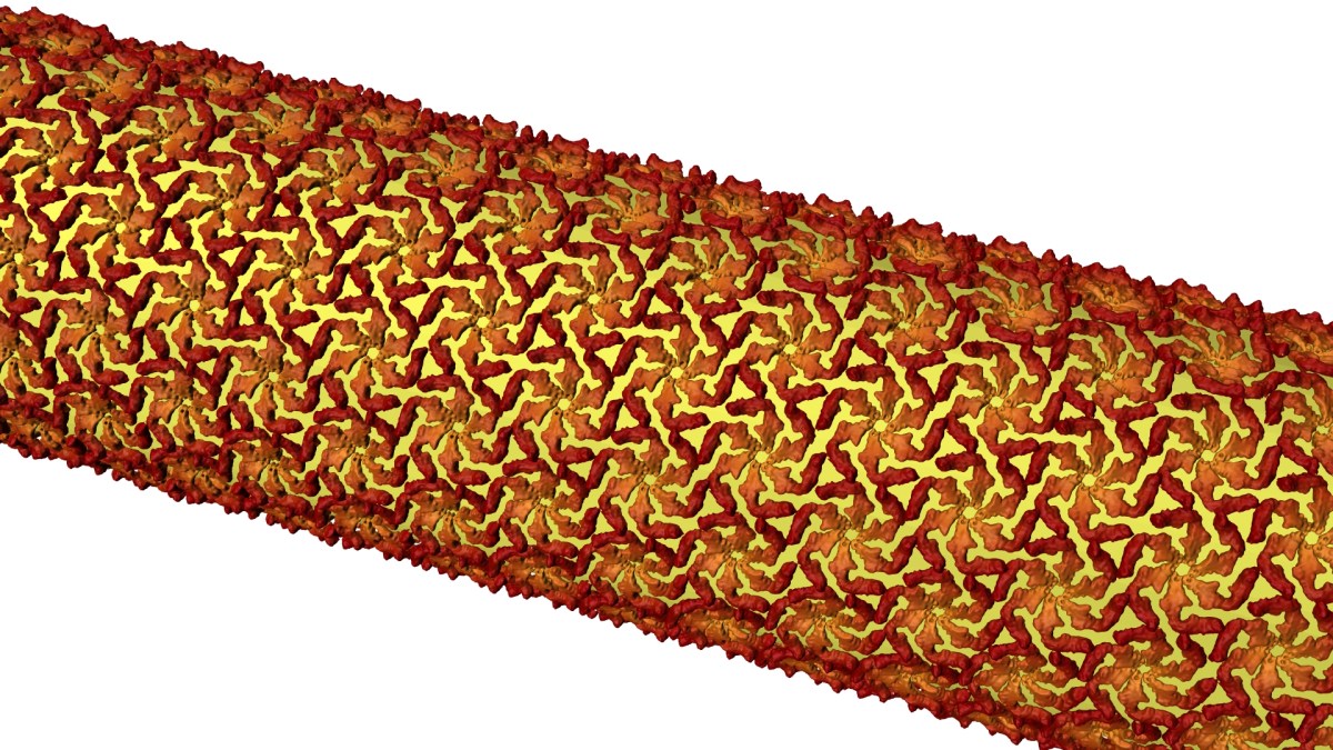
Then they tracked the glowing building blocks with single-molecule microscopy as they formed a shell that covered the microbe in a hexagonal, tile-like pattern (shown in image above) in less than two hours. A technique called stimulated emission depletion (STED) microscopy allowed them to see structural details of the layer as small as 60 to 70 nanometers, or billionths of a meter, across – about one-thousandth the width of a human hair.”

The scientists were surprised by what they saw: the protein molecules spontaneously assembled themselves without the help of enzymes.

“It’s like watching a pile of bricks self-assemble into a two-story house,” said Jonathan Herrmann, a graduate student in structural biology at Stanford involved in the study, in the news release.

The researchers believe the protein building blocks are guided to form in specific regions of the cell surface by small defects and gaps within the S-layer. These naturally-occurring defects are inevitable because the flat crystalline sheet is trying to cover the constantly changing, three-dimensional shape of the bacterium, they said.

Among other applications, they hope their findings will offer potential new targets for drug treatments.

“Now that we know how they assemble, we can modify their properties so they can do specific types of work, like forming new types of hybrid materials or attacking biomedical problems,” said Soichi Wakatsuki, PhD, a professor of structural biology and photon science at SLAC, in the release.

Illustration by Greg Stewart/SLAC National Accelerator Laboratory

This is a reposting of my Scope blog story, courtesy of Stanford School of Medicine.

Predicting women at risk of preeclampsia before clinical symptoms

Many of my female friends became pregnant with their first child in their late 30s or early 40s, which increased their risk of common complications such as high blood pressure, gestational diabetes and preeclampsia.

Affecting over 8 million women worldwide, preeclampsia can lead to serious, even fatal, complications for both the mother and baby. The clinical symptoms of preeclampsia typically start at 20 weeks of pregnancy and include high blood pressure and signs of kidney or liver damage.

“Once these clinical symptoms appear, irreparable harm to the mother or the fetus may have already occurred,” said Stanford immunologist Brice Gaudilliere, MD, PhD.  “The only available diagnostic blood test for preeclampsia is a proteomic test that measures a ratio of two proteins. While this test is good at ruling out preeclampsia once clinical symptoms have occurred, it has a poor positive predictive value.”

Now, Stanford researchers are working to develop a diagnostic blood test that can accurately predict preeclampsia prior to the onset of clinical symptoms.

A new study conducted at Stanford was led by senior authors Gaudilliere, statistical innovator Nima Aghaeepour, PhD, and clinical trial specialist Martin Angst, MD, and co-first authors and postdoctoral fellows Xiaoyuan Han, PhD, and Sajjad Ghaemi, PhD. Their results were recently published in Frontiers in Immunology.

They analyzed blood samples from 11 women who developed preeclampsia and 12 women with normal blood pressure during pregnancy. These samples were obtained at two timepoints, allowing the scientists to measure how immune cells behaved over time during pregnancy.

“Unlike prior studies that typically assessed just a few select immune cell types in the blood at a single timepoint during pregnancy, our study focused on immune cell dynamics,” Gaudilliere explained. “We utilized a powerful method called mass cytometry, which measured the distribution and functional behavior of virtually all immune cell types present in the blood samples.”

The team identified a set of eight immune cell responses that accurately predicted which of the women would develop preeclampsia — typically 13 weeks before clinical diagnosis.

At the top of their list was a signaling protein called STAT5. They observed higher activity of STAT5 in CD4+ T-cells, which help regulate the immune system, at the beginning of pregnancy for all but one patient who developed preeclampsia.

“Pregnancy is an amazing immunological phenomenon where the mother’s immune system ‘tolerates’ the fetus, a foreign entity, for nine months,” said Angst. “Our findings are consistent with past studies that found preeclampsia to be associated with increased inflammation and decreased immune tolerance towards the fetus.”

Although their results are encouraging, more research is needed before translating them to the clinic.

The authors explained that mass cytometry is a great tool to find the “needle in the haystack.” It allowed them to survey the entire immune system and identify the key elements that could predict preeclampsia, but it is an exploratory platform not suitable for the clinic, they said.

“Now that we have identified the elements of a diagnostic immunoassay, we can use conventional instruments such as those used in the clinic to measure them in a patient’s blood sample.” Aghaeepour said.

First though, the team needs to validate their findings in a large, multi-center study. They are also using machine learning to develop a “multiomics” model that integrates these mass cytometry measurements with other biological analysis approaches. And they are investigating how to objectively define different subtypes of preeclampsia.

Their goal is to accurately diagnose preeclampsia before the onset of clinical symptoms.

 “Diagnosing preeclampsia early would help ensure that patients at highest risk have access to health care facilities, are evaluated more frequently by obstetricians specialized in high-risk pregnancies and receive treatment,” said Gaudilliere.

Women with preeclampsia can receive care through the obstetric clinic at Lucile Packard Children’s Hospital Stanford.

Photo by Pilirodriquez

This is a reposting of my Scope blog story, courtesy of Stanford School of Medicine.

Explaining neuroscience in ongoing Instagram video series: A Q&A

At the beginning of the year, Stanford neuroscientist Andrew Huberman, PhD, pledged to post on Instagram one-minute educational videos about neuroscience for an entire year. Since a third of his regular followers come from Spanish-speaking countries, he posts them in both English and Spanish. We spoke soon after he launched the project. And now that half the year is over, I checked in with him about his New Year’s resolution.

How is your Instagram project going?

“It’s going great. I haven’t kept up with the frequency of posts that I initially set out to do, but it’s been relatively steady. The account has grown to about 13,500 followers and there is a lot of engagement. They ask great questions and the vast majority of comments indicate to me that people understand and appreciate the content. I’m really grateful for my followers. Everyone’s time is valuable and the fact that they comment and seem to enjoy the content is gratifying.”

What have you learned?

“The feedback informed me that 60 seconds of information is a lot for some people, especially if the topic requires new terms. That was surprising. So I have opted to do shorter 45-second videos and those get double or more views and reposts. I also have started posting images and videos of brains and such with ‘voice over’ content. It’s more work to produce, but people seem to like that more than the ‘professor talking’ videos.

I still get the ‘you need to blink more!’ comments, but fortunately that has tapered off. My Spanish is also getting better but I’m still not fluent. Neural plasticity takes time but I’ll get there.”

What is your favorite video so far?

“People naturally like the videos that provide something actionable for their health and well-being. The brief series on light and circadian rhythms was especially popular, as well as the one on how looking at the blue light from your cell phone in the middle of the night can potentially alter sleep and mood. I particularly enjoyed making that post since it combined vision science and mental health, which is one of my lab’s main focuses.”

What are you planning for the rest of the year?

“I’m kicking off some longer content through the Instagram TV format, which will allow people who want more in-depth information to get that. I’m also helping The Society for Neuroscience get their message out about their annual meeting. Other than that, I’m just going to keep grinding away at delivering what I think is interesting neuroscience to people that would otherwise not hear about it.”

Is it fun or an obligation at this point?

“There are days where other things take priority of course — research, teaching and caring for my bulldog Costello — but I have to do it anyway since I promised I’d post. However, it’s always fun once I get started. If only I could get Costello to fill in for me when I get busy…”

This is a reposting of my Scope story, courtesy of Stanford School of Medicine.

Simplified analysis method could lead to improved prosthetics, a Stanford study suggests

Brain-machine interfaces (BMI) are an emerging field at the intersection of neuroscience and engineering that may improve the quality of life for amputees and individuals with paralysis. These patients are unable to get signals from their motor cortex — the part of the brain that normally controls movement — to their muscles.

Researchers are overcoming this disconnect by implanting in the brain small electrode arrays, which measure and decode the electrical activity of neurons in the motor cortex. The sensors’ electrical signals are transmitted via a cable to a computer and then translated into commands that control a computer cursor or prosthetic limb. Someday, scientists also hope to eliminate the cable, using wireless brain sensors to control prosthetics.

In order to realize this dream, however, they need to improve both the brain sensors and the algorithms used to decode the neural signals. Stanford electrical engineer Krishna Shenoy, PhD, and his collaborators are tackling this algorithm challenge, as described in a recent paper in Neuron.

Currently, most neuroscientists process their BMI data looking for “spikes” of electrical activity from individual neurons. But this process requires time-consuming manual or computationally-intense automatic data sorting, which are both prone to errors.

Manual data sorting will also become unrealistic for future technologies, which are expected to record thousands to millions of electrode channels compared to the several hundred channels recorded by today’s state-of-the-art sensors. For example, a dataset composed of 1,000 channels could take over 100 hours to hand sort, the paper says. In addition, neuroscientists would like to measure a greater brain volume for longer durations.

So, how can they decode all of this data?

Shenoy suggests simplifying the data analysis by eliminating spike sorting for applications that depend on the activity of neural populations rather than single neurons — such as brain-machine interfaces for prosthetics.

In their new study, the Stanford team investigated whether eliminating this spike sorting step distorted BMI data. Turning to statistics, they developed an analysis method that retains accuracy while extracting information from groups rather than individual neurons. Using experimental data from three previous animal studies, they demonstrated that their algorithms could accurately decode neural activity with minimal distortion — even when each BMI electrode channel measured several neurons. They also validated these experimental results with theory.

 “This study has a bit of a hopeful message in that observing activity in the brain turns out to be easier than we initially expected,” says Shenoy in a recent Stanford Engineering news release. The researchers hope their work will guide the design and use of new low-power, higher-density devices for clinical applications since their simplified analysis method reduces the storage and processing requirements.

This is a reposting of my Scope blog story, courtesy of Stanford School of Medicine.

Photo by geralt.