Building off-the-shelf Lego robots can teach kids important skills like mechanical engineering, computer programming and teamwork. Now, Stanford bioengineers are adding life sciences and chemistry to the list.
Stanford researchers have developed a liquid-handling Lego robot capable of a range of experiments — integrating robotics, biology, chemistry, programming and hands-on learning into a single, open-source educational tool. Built from a cheap plastic syringe and a Lego Mindstorm EV3 Education kit, the robots are designed to pipette fluids into and out of plastic containers commonly used in laboratories.
The team also designed and tested several fundamental experiments for elementary and middle school students using their DIY robots and common household items like food coloring, salt or sugar, which are described in a recent paper published in PLOS Biology.
One of the favorites is an experiment that teaches kids about density and buoyancy by sequentially layering colored liquids with different salt concentrations into a single test tube — demonstrating that the liquids float on top of each other instead of mixing, and explaining why objects float or sink.
“We would love it if more students, do-it-yourself learners, STEM teachers and researchers would embrace this type of work, get excited and then develop additional open-source instructions and lesson plans for others to use,” said Ingmar Riedel-Krus. PhD, assistant professor of bioengineering, in a recent news release.
This is a reposting of my Scope blog story, courtesy of Stanford School of Medicine.
What do immersive simulations, filmmaking and emergency medicine have in common? One answer is Henry Curtis, MD, a Stanford clinical instructor in emergency medicine who’s using innovative tools to educate medical students and residents about emergency medicine.
Curtis’s latest endeavor is a class called EMED 228: Emergency Video Production, which teaches students how to impact emergency care through film by “telling a story that matters.” I recently spoke with him about his use of filmmaking and simulation games.
Why do you use simulations and filmmaking as education tools?
“Both simulation and filmmaking serve different purposes for emergency medicine education. Immersive simulation is an arena. It’s a place where learners can experience a medical emergency in a safe environment. They make medical decisions, perform procedures and communicate with the patient and their team. When it is all over, they reflect on what happened. Aside from real life clinical experience, there is no better educational technique.
Filmmaking imagines and documents life. Video based learning has many advantages, not the least of which is reproducibility —a final cut is independent of individual human factors that could affect quality on any given day. It is fascinating to bind the experiences unfolding in a simulated medical emergency with videos. For instance, engagement videos can function to more powerfully immerse the learner into a given clinical scenario. Information videos can relate valuable educational cues more effectively than a photo, announcement or text flashed on a screen. Video based debriefing allows playback of the important moments in a scenario.”
What inspired you to create the EMED 228 course? What does it entail?
“I’ve been pursuing a master of fine arts in directing for the last few years at the Academy of Art University in San Francisco. I wanted to give back to Stanford and share the filmmaking skills I’ve acquired with students.
EMED 228 is open to undergrads, grads and medical students. We were fortunate to have a nice mix of students of all different educational interests and filmmaking experience enrolled. They were exposed to an overview of filmmaking. We began the first day with theory. The class then quickly progressed to understanding and implementing the practical aspects of creating a final product — using a robust array of equipment, including multiple high-definition DSLR cameras, GoPros, drones, remote focus pulling devices and gimbals.
The entire class culminated in a screening and Q&A session of the documentary that we created titled, Care Flight in the Golden Hour. We aimed to provide insight into the process and people delivering care to critically ill patients in Lake Tahoe requiring air medical evacuation. These caregivers provide a service, which oftentimes will make the difference between life and death of healthy people who are having a tragic day. We chose to film on location in Truckee, California. Hannah Rasmussen, a first-year medical student, acted as a teaching assistant. Her efforts were invaluable in organizing our remote and on-site collaborations.”
As a child, did you want to be a film director when you grew up?
“I did not always know that I would be so drawn to the storytelling art of filmmaking or that I would prefer to be in the role of directing. I did know that I preferred film to photos when creating memories. In fact, I have many more short videos than photos in the memory closet. During the last year of my emergency medicine residency, I chose to concentrate on the use of film in disaster medicine education and this is where my filmmaking life really began.
Stanford University is a rich world of opportunity. It has encouraged me to chase my interests and carve out a niche in the medical humanities. The department of emergency medicine is fully supportive of my journey. With such resources and encouragement offered at so many levels, I encourage everyone to seek out their passion in this environment.”
This is a reposting of my Scope blog story, courtesy of Stanford School of Medicine.
Breast cancer patients are often faced with a difficult decision at the end of their primary treatment: Should they get systemic adjuvant therapy, such as the anti-estrogen drug tamoxifen? Such therapies lower the risk that the cancer will come back, but they also carry the risk of potentially serious side effects.
What would be helpful is for physicians to have a way to predict which patients have the best prognosis and might not need adjuvant therapy. Now, researchers from the Lawrence Berkeley National Laboratory may have a solution, according to a study recently published in Oncotarget.
The research team analyzed clinical patient data and large genomic datasets of normal and tumor breast tissues — identifying 381 genes associated with the relapse-free survival of breast cancer patients. With further analysis, they were able to develop a scoring system based on a 12-gene signature that predicts breast cancer survival. Patients with a low score were more likely to live longer.
“Distinguishing patients with good prognosis could potentially spare them the toxic side effects associated with adjuvant therapy. Determining prognosis involves a range of other clinical factors, including tumor size and grade, the degree to which the cancer has spread, and the age and race of the patient. Our scoring system was predictive of survival independent of these other variables.”
The study showed that their 12-gene signature was effective at predicting patient survival for two specific subtypes of breast cancer — luminal-A and HER2 — but it wasn’t effective for other subtypes.
In addition, the researchers identified seven genes as potential tumor suppressors that could be targeted when developing new breast cancer therapies. They hope that their work will help doctors and patients make more informed treatment decisions, as well as help others develop better breast cancer drugs.
This is a reposting of my Scope blog story, courtesy of Stanford School of Medicine.
Will people read about null research findings? And are such findings news? These are critical questions facing health reporters, because news coverage often influences how people make their health-care decisions.
The problem is that positive research findings make more alluring stories, particularly if the new study suggests a potential cure for a horrible disease. But many of these initially positive findings are refuted by larger, more rigorous follow-up studies that journalists rarely cover. This biased news coverage can mislead the public with important consequences — such as helping to perpetuate the discredited link between autism and vaccines.
Health News Review recently tackled this topic, using the example of statins. Initial observational studies showed that statins may help boost survival from cancer. But later, more rigorous trials showed that statins don’t improve cancer outcomes. Nonetheless, the media heavily covered the initial findings, but barely picked up on the more reliable negative findings.
Researchers at the University of Bordeaux, France investigated the extent of this problem by analyzing the news coverage of 156 primary medical studies, as outlined in a paper recently published in PLOS One. They focused on studies that looked for associations between risk factors and diseases in six areas: psychiatry, neurology, breast cancer, rheumatoid arthritis, glaucoma and psoriasis.
The study found that “journalists preferentially cover initial findings although they are often contradicted by meta-analyses and rarely inform the public when they are disconfirmed.”
Using a database of thousands of stories published in the general press, the research team discovered all 53 initial research studies that generated news coverage reported positive findings — even though two thirds of these initial findings were refuted by subsequent research. In contrast, journalists covered none of the 174 initial studies reporting a null effect and rarely covered null findings in subsequent studies.
They also found that journalists more often covered lifestyle research, which investigates factors like diet and smoking. Lifestyle associations received larger newspaper coverage, even if the initial studies were published in less prestigious scientific journals. The authors stated, “This preferential coverage further supports the view that the first journalists’ aim is to attract readers’ attention.”
Finally, the researchers offered some advice on how to remedy this problem. They suggested that “journalists should always ask scientists whether it is an initial finding and, if so, they should inform the public that this discovery is still tentative and must be validated by subsequent studies.” They also recognized that it can be difficult for journalists to find objective sources to put a new study into the appropriate context, so they called on scientists to assist journalists. The authors concluded by saying that scientists have a moral duty to make sure press releases covering their work are accurate.
This is a reposting of my Scope blog story, courtesy of Stanford School of Medicine.
Traumatic brain injuries, like those caused by concussions, are common. But suffering even a mild brain injury boosts the likelihood of developing neurological and psychiatric disorders, such as Alzheimer’s disease and posttraumatic stress disorder, years later. Exactly how and why that happens remains a mystery.
“Very little is known about how people with brain trauma — like football players and soldiers — develop neurological disorders later in life,” said Fernando Gomez-Pinilla, PhD, a University of California, Los Angeles professor of neurosurgery and of integrative biology and physiology, in a recent news release.
Now, Gomez-Pinilla and his colleagues have discovered that a brain injury harms “master” genes that control other genes throughout the body. This triggers the alteration of hundreds of genes, which are linked to disorders like Alzheimer’s disease, Parkinson’s disease, PTSD, attention deficit hyperactivity disorder and depression. Their study was recently published in EBioMedicine.
In the study, the researchers trained 20 rats to navigate through a maze. They then injected a fluid into the brain of half the rats to simulate a concussion-like brain injury. When all the rats were retested in the maze, the rats with a brain injury took about 25 percent longer than the controls to solve the maze — indicating a change in basic cognitive function.
Next, the team investigated how the brain injuries altered the rats’ genes. They analyzed RNA samples from the rats’ white blood cells and hippocampi, the part of the brain that plays a central role in memory processes. In the injured rats, they found almost 300 genes had been altered in the hippocampus and over 1200 genes in the white blood cells.
More than 100 of these altered genes have counterparts in humans that are linked to neurological and psychiatric disorders. The researchers concluded that concussive brain injury reprograms key genes and this reprogramming could make neurological and psychiatric disorders more likely.
In addition, almost two dozen of the altered genes occurred in both the hippocampus and white blood cells. The researchers hope this genetic signature can be used to develop a gene-based blood test that determines whether a brain injury has occurred and whether future neurological disorders are likely.
They also hope their identification of master genes can give scientists new targets to develop better pharmaceuticals for brain disorders. However, more research is needed to fully understand the role of these master genes. Gomez-Pinilla said he now plans to study the phenomenon in people who have suffered a traumatic brain injury.
This is a reposing of my Scope blog story, courtesy of Stanford School of Medicine.
How often, in the last two weeks, have you felt tired or lacked energy?
Daily? Never? For me, and I’m guessing for many of you, the answer is somewhere in between.
Researchers posed that question to tens of thousands of study participants to investigate whether tiredness has a genetic basis. They found that genes play a small but significant role in overall fatigue.
The multi-institutional team of researchers analyzed genetic data from the UK Biobank for 108,976 individuals who reported whether they had felt tired in the last two weeks. The participants selected four possible answers, ranging from “not at all” to “nearly every day”; most answered either “not at all” or “several days.”
The researchers found that genetic factors account for about 8 percent of the participants’ differences in self-reported tiredness, according to a paper recently published in Molecular Psychiatry. This implies that tiredness is largely due to other factors, such as not getting enough sleep.
Some inherent factors such as personality traits or poor health can contribute, however. By averaging tiredness across a large sample and performing a genomic-wide association study, the researchers identified genetic links between tiredness and inherent factors — using the UK Biobank’s data on the participants’ physical health, mental health, personality and cognitive functioning.
They found that an individual’s genetic predisposition to some physical and mental illnesses — not just the presence of these illnesses — was associated with feeling tired. For instance, people who were genetically prone to Type 2 diabetes were also prone to tiredness, even if they did not have diabetes.
The authors summarized that tiredness is a “partly heritable, heterogeneous and complex phenomenon,” which requires further research to fully understand. However, they indicate that most people’s differences in tiredness can be attributed to external factors such as the lack of sleep.
This is a reposting of my Scope blog story, courtesy of Stanford School of Medicine.
When I think about bicycle safety, I think of helmets, lights and strategies to share the road with cars.
But physicians have a different perspective — too many hours on a bicycle saddle can compress vital arteries and nerves to cause numbness, pain and sexual dysfunction. This risk is likely affected by the design of the saddle, fit of the bike, riding position, ride duration and a host of other factors.
But there’s a lot that remains unknown. So Michael Eisenberg, MD, an assistant professor of urology at Stanford, is conducting the Stanford CYCling and Lower Effects (CYCLE) study to hone in on the factors affecting the comfort and safety of cycling. He’s collaborating with Roger Minkow, MD, a Bay Area-based saddle designer and ergonomic consultant.
The researchers are inviting volunteers to answer a brief online survey about their bicycling habits, equipment and health. I recently reached out to Eisenberg to learn more.
What inspired you to conduct the CYCLE study?
“About 20 years ago, several studies demonstrated an association between cycling, erectile dysfunction and even infertility. Many of these health issues can be reversed if caught early, but they can become permanent over time. Since then, the bicycle industry has undergone a major redesign of equipment to try to mitigate the risk. And it’s been years since a large study has been conducted to understand the current prevalence of sexual dysfunction in riders and to understand if there are cycling related factors — such as duration of riding and saddle design — that are contributing.
Cycling is quite popular in this area and I have several patients who have come in over the years complaining of genital pain, numbness or performance issues. Recently, the saddle designer Roger Minkow reached out to me about the topic. We created and initiated the CYCLE study in October 2016 to help understand the current state of cycling on pelvic and sexual health for male and female riders. We’re very excited about the study and hope it will help make the sport safer and more comfortable.”
How can cyclists participate?
“Cyclists participate in the study by completing a brief online survey that takes about 15 minutes. In the survey, we obtain a comprehensive look at the cycling habits of men and women, including the type of riding they do, their intensity level and details about their equipment. We then ask participants about their overall health — such as their weight, body measurements and basic medical history. Finally, we ask validated questions related to sexual function and how it corresponds to their riding habits.
Nearly 1500 people have participated in the study so far. There are so many different types of cyclists and equipment in common use. In order for us to effectively compare these, we need about 8500 more participants.”
Can you give an example of how you treat a cycling-related health problem?
“I recently saw a man with persistent penile numbness after several long bike rides. We reviewed risk factors and pelvic anatomy related to his condition. We then discussed certain cycle practices he can modify to allow him to be able to cycle as much as he’d like without the symptoms, and these modified practices have worked well.
In generally, cyclists really love to ride so my goal is not to tell them to stop. I look at a patient’s equipment, body position, saddle design, riding habits, and when symptoms occur — to come up with a personalized strategy for that rider. In select cases, I even prescribe some medications to help circulation.”
Do you cycle?
“Yes, I cycle on the road for both pleasure and exercise. We live in a beautiful area. One of my favorite rides is around my neighborhood along Foothill Expressway and Junipero Serra Boulevard.”
This is a reposting of my Scope blog story, courtesy of Stanford School of Medicine.
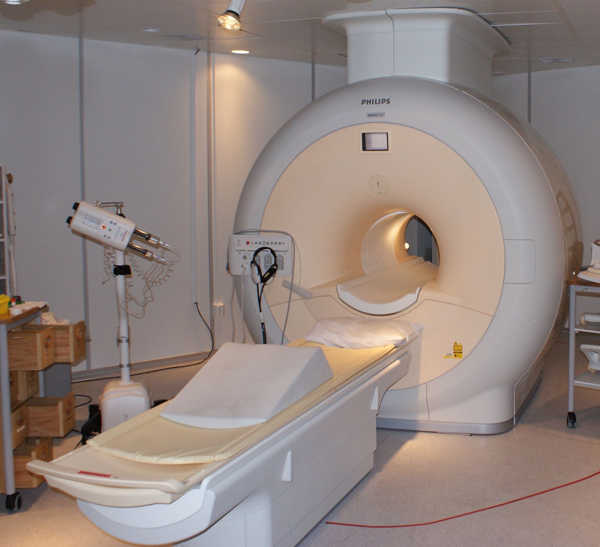
MRI is a powerful, non-invasive diagnostic tool widely used to investigate anatomical structures and functions in the body.
Though generally considered to be safe, several studies in the last decade have reported an increase in DNA damage, or genotoxicity, due to cardiac MRI scans. Other research doesn’t support these findings — raising the controversial question of whether an MRI’s electromagnetic fields pose a health threat.
A multi-institutional research team explored this issue by reviewing the literature published between 2007 and 2016. Specifically, the group considered three questions during their review:
Do MRIs really cause genotoxicity?
What are the potential adverse health effects of exposure to MRI electromagnetic fields?
What impact does this have on patient health?
As outlined in a commentary appearing in Radiation Research, the evidence correlating MRIs with genotoxicity “is, at best, mixed.” After emphasizing the limitations of existing studies, which typically included at most 20 participants and lacked sufficient quality control measures, the authors summarized:
“We conclude that while a few studies raise the possibility that MRI exams can damage a patient’s DNA, they are not sufficient to establish such effects, let alone any health risk to patients. … We consider that genotoxic effects of MRI are highly unlikely.”
A previous 2015 review paper published in Mutation Research called for comprehensive, international, multi-centered collaborative studies to address this issue, using a common and widely used MRI exposure protocol with a large number of patients.
The authors of the new review note that such studies would be very expensive and would require hundreds of thousands of participants, which may not be warranted.
“If you want to do something next, do a very well-designed, large study of the types that have already been done, but with better statistics and better controls,” said John Moulder, PhD, a professor emeritus at the Medical College of Wisconsin, in a recent news story. “And make sure that this punitive genotoxicity is even real before beginning more expensive follow-up studies.”
This is a reposting of my Scope blog story, courtesy of Stanford School of Medicine.


