One researcher’s journey to understand the molecular basis of aging, using blood

Image by Cliker-Free-Vector-Images

Hanadie Yousef, PhD, studies how the body ages — first as a graduate student at UC Berkeley and currently as a postdoctoral research fellow at Stanford with her mentor, Tony Wyss-Coray, PhD, professor of neurology.

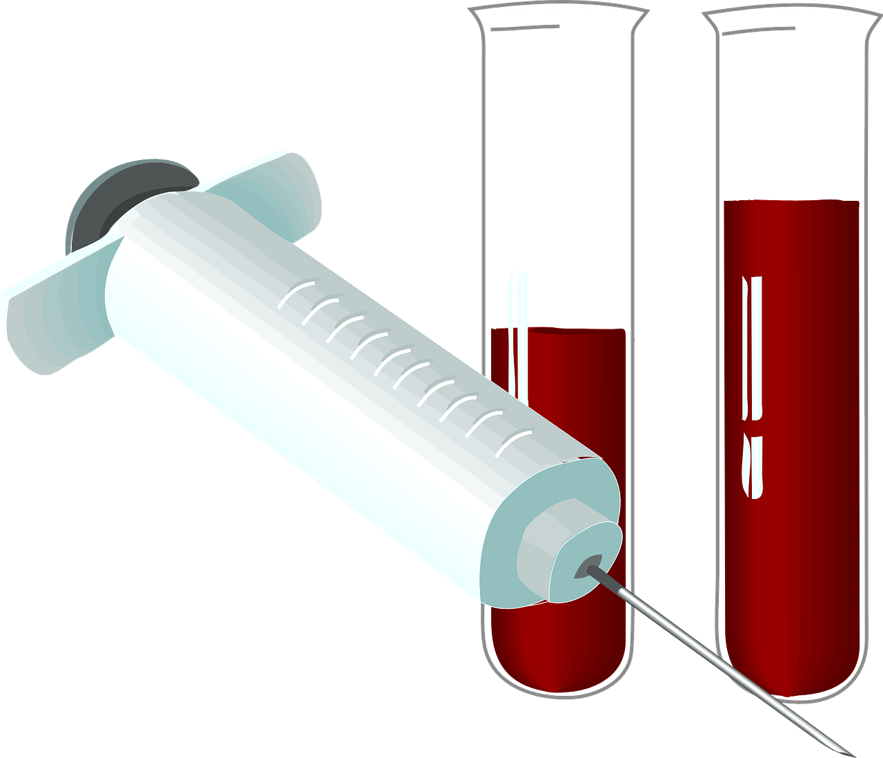
Her current anti-aging research is based on a pivotal study performed at Stanford in the laboratory of Thomas Rando, MD, PhD, professor of neurology and neurological sciences. In this 2005 study, researchers surgically connected the circulatory systems of a young and old mouse. Within a few weeks, the young mouse’s blood began rejuvenating the tissues in the old mouse. Specifically, the young blood rejuvenated organ stem cells in the brain, muscle and liver of the old mouse. When activated, these adult stem cells fend off aging by replenishing depleted cells and regenerating damaged tissues. 

Subsequent studies by the Wyss-Coray lab and others have shown that organ stem cells retain their regenerative capacity, but the biochemical cues that control their function change with age — causing the abandonment of tissue maintenance and repair in the elderly. This can be overcome by injecting young blood to enhance the adult stem cells’ environment.

“In graduate school, I became fascinated with the idea that adult tissues could be rejuvenated by their tissue-specific stem cells, which severely decline in function as we grow old,” said Yousef. “So I wanted to understand the molecular mechanisms underlying this decline.”

Specifically, Yousef examined a molecule called transforming growth factor-beta 1 (TGF-β1). “In young people, TGF-β1 exists at lower levels around stem cells and helps regulate tissue regeneration. However, elevated levels of TGF-β1 in the stem cell microenvironment in older people promotes inflammation and prevents stem cell activity, causing decline in tissue function,” Yousef said.

As a graduate student, to help find a potential treatment, she tested a cancer drug called Alk-5 kinase inhibitor, which decreased the levels of TGF-β1 signaling to stem cells, increasing stem cell activity —when systematically injected into aged mice.

“The control old mice had extensive scar tissue following muscle injury, while the old mice that got the Alk-5 inhibitor were systematically able to generate new muscle fibers almost as well as young animals,” Yousef said. “Additionally, we saw that the inhibitor could cross the blood brain barrier and act locally on neural stem cells to enhance the formation of new adult neurons in the hippocampus, the part of the brain important for learning and memory function that severely declines with age.”

The Berkeley scientists are now considering ways to evaluate the drug’s efficacy in humans. The hope is to be able to inject a drug, like the Alk-5 inhibitor, to prevent the onset of multiple diseases associated with aging, including Alzheimer’s.

And Yousef continues her anti-aging research at Stanford. “I now study the opposite effect: what does old blood do to young brain function?” Yousef said. “We’ve seen that aged blood plasma is more pro-inflammatory than young blood plasma, and it contributes to increased brain inflammation and inhibited neural stem cell function with aging.”

So young blood improves brain function, whereas old blood impairs it. Does this mean that young blood can be injected into older patients to treat degenerative brain disorders like Alzheimer’s? According to Yousef, the answer is:

Potentially, yes! In our lab, my colleagues have shown that young blood can improve brain function in Alzheimer’s mice models, and their results will be published soon. Based on these studies, there is an ongoing clinical trial at Stanford, called the Plasma for Alzheimer’s Symptom Amelioration study, in which Alzheimer’s patients receive young blood transfusions to see if this can reduce the severity of disease progression.

A lot of research is needed before a successful treatment is found for Alzheimer’s, but understanding the rejuvenating factors in young blood may be a key step. “We’re working intensively to find out what those factors might be and from exactly which tissues they originate,” said Wyss-Coray in an earlier news release.

This is a reposting of my Scope blog story, courtesy of Stanford School of Medicine.

Most primary care doctors have favorite patients, study finds

Image by Regina Holliday

As a teacher, I try to treat all my students equally but I definitely have favorites. I’m sure other teachers have favorite students too, so it makes sense to learn that physicians have favorite patients.

A team of researchers led by Joy Lee, PhD, a postdoctoral fellow at the Johns Hopkins Bloomberg School of Public Health, interviewed 25 primary care physicians who worked in clinical settings within the Johns Hopkins medical system about their favorite patients. The participating physicians were predominantly white and about evenly split between male and female.

The doctors were a bit uncomfortable with the term ‘favorite patient,’ the researchers reported in Patient Education and Counseling. It raised concerns regarding boundaries and favoritism. However, all but three of the participating physicians admitted to having favorite patients.

The goal of the study was to identify the common attributes of these physicians’ favorite patients and examine how having favorite patients impacts their physician-patient relationships.

Who were the favorites? Surprisingly, they weren’t typically the most compliant patients or the ones most similar to the doctors. Instead, they were long-term patients who spent more time with their physician while going through a major illness. So the doctors were very familiar with their favorite patients’ personalities and health histories — allowing them to provide the best care.

“For patients, these findings highlight the importance of having a usual source of care, a primary care doctor with whom they can establish a relationship,” said Lee in a recent news release. “Favorite patients might not be consistently sick, but when a crisis comes they have an existing relationship to work off of.”

Of course having favorites isn’t the same as playing favorites. The participating physicians argued that their awareness of having both favorite and challenging patients helps them prevent favoring the care of certain patients over others. They also generally like most of their patients.

“This concern demonstrates that physicians are striving to be fair and to give all their patients the best possible care,” Lee said. “We discovered that doctors really thought about their relationship with patients, which is encouraging from a patient perspective. Their thinking really humanizes the patient-physician relationship.”

This is a reposting of my Scope blog story, courtesy of Stanford School of Medicine.

Trojan Horses: Nanoparticles sneak drugs into brain to battle cancer

Photo by dierk schaefer

I just read an interesting article in the Berkeley Science Review about using nanoparticles to make chemotherapy more effective against a type of brain cancer called glioblastoma. I was then surprised and proud when I realized one my former science-writing students, Dharsi Devendran, wrote it.

Although rare, glioblastoma is an invasive and deadly brain cancer with octopus-like tentacles that are difficult to completely remove with surgery. Even the standard combined treatment of surgery, radiation and chemotherapy isn’t very effective — people typically die within months of diagnosis. So researchers are actively searching for better treatments.

Devendran explains:

Although it is difficult to fight, glioblastoma also has a weakness. In its rush to feed itself, it accelerates the blood vessel formation process and creates hole-riddled blood vessels around the tumor. Because cancer drugs are small enough to slip through these holes, they can exploit this defect—but they also need a strategy to cross the blood-brain barrier in order to reach the tumor.

Acting like a security system for the brain, the blood-brain barrier is a network of blood vessels that allow essential nutrients to enter while blocking harmful molecules. Unfortunately, it also blocks life-saving chemotherapy drugs, unless researchers can find clever ways to sneak them through the barrier.

Ting Xu, PhD, professor of materials science at UC Berkeley, and her collaborators are developing tiny nanocarriers that can envelop and protect chemotherapy drugs as they move through the blood, across the blood-brain barrier, and into the brain to the glioblastoma tumor tissue.

The researchers designed a new nanocarrier, called a 3-helix micelle (3HM), out of proteins with molecules on the surface that fit into specific proteins found only on the surface of the tumor cells — like fitting a key into a lock. Once the 3HM nanocarriers access the tumor cells, they release their chemotherapy drugs to help destroy the glioblastoma.

Xu’s team has shown that their 3HM nanocarrier is twice as effective at reaching glioblastoma cells as liposomes, a nanocarrier made of fatty acids that is a standard in nanotechnology drug delivery. This is in part because the 3HM is five times smaller than liposomes.

“We still don’t understand the mechanism completely,” said JooChuan Ang, a graduate student in Xu’s group, in the article. “Size is definitely a factor, but there could be other factors that contribute…”

This is a reposting of my Scope blog story, courtesy of Stanford School of Medicine.

Quitting smoking: Best drug differs for men and women

Papierosa_1_ubt_0069
Photo by Tomasz Sienicki

It’s tough to quit smoking. I’ve seen friends and family members struggle with nicotine withdrawal symptoms: cravings for tobacco, anxiety, anger, irritation, increased hunger and even trouble thinking.

One out of six adults in the United States currently smoke and about half of them are trying to quit, but the success rate remains low. What’s the best way to stop smoking? A new study may help point the way — for women.

The study found that a medication frequently used to help smokers quit is more effective for women than men. Philip Smith, PhD, assistant medical professor at the City College of New York, led the multi-institutional study: a network meta-analysis of 28 randomized clinical trials involving a total of 14,389 smokers (51 percent female).

The researchers did a head-to-head comparison between the three common types of medications used for smoking cessation: the nicotine patch, varenicline (sold as Chantix and Champix) and sustained-release bupropion (sold as Wellburtin or Zyban). The quit rate of the participants was based on biochemical verification of their abstinence after six months.

The authors reported in their new paper in Nicotine & Tobacco Research:

“Women treated with varenicline were 41 percent more likely to achieve 6-month abstinence compared to women treated with TN [transdermal nicotine patch], and were 38 percent more likely to achieve 6-month abstinence than women treated with bupropion. For men, the benefit of varenicline over TN and bupropion were smaller and were not statistically significant.”

“Before our study, research had shown that among the choices for medications for smokers who wanted to quit, varenicline was the clear winner when it came to promoting quitting,” said Smith in a recent news release. “Our study shows this is clearly the case for women. The story seems less clear among men, who showed less of a difference when taking any of the three medications.”

The research findings identify varenicline as a particularly potent first option treatment for women. However, the good news for all smokers is that all three medicines significantly improved quit rates for both men and women, when compared with placebo.

If you’re trying to quit smoking, a combination of counseling and medication has been shown to be an effective way to treat tobacco dependence — speak with your doctor or contact a smoking cessation program.

This is a reposting of my Scope blog story, courtesy of Stanford School of Medicine.

From art to surgery: Stanford alumna reconstructs new ears for children

Dr Sheryl Lewin in the operating room (courtesy of Lewin)

Some children are born with a missing or malformed small ear due to a rare congenital condition called microtia. In most cases, the child’s ear canal is also very small or absent, resulting in hearing loss.

The surgical procedures used to correct microtia require the skills of both a sculptor and surgeon — making it the perfect specialty for Sheryl Lewin, MD, a craniofacial plastic surgeon who began her training as an artist and architect.

Lewin’s career has been passionately devoted to treating microtia through her private medical practice and nonprofit organization called Earicles, which helps children born without ears through education, research and free or reduced-cost treatment. I recently spoke with Lewin about her work: 

As an architect major, what inspired you to become a physician?

“When I was in architecture school at UC Berkeley, I loved the challenge of design, where you can use your own creativity to solve visual and spatial problems. My program was heavily artistic — we drew, painted and sculpted. But what was missing was the ability to use those skills to directly affect someone’s life in a tangible and meaningful way.

During college, I lived across the street from an elementary school that served underprivileged kids, which inspired me to start a volunteer organization of Berkeley undergraduates that mentored disadvantaged children in the local community. I recognized that I really enjoyed working and helping kids, and medicine was a way to do that.

When I went to medical school at Stanford, I was drawn to surgery as it gave me the ability to work with my hands. I decided to pursue pediatric plastic surgery after I saw my first cleft lip surgery on a tiny infant, whose life was transformed in a couple of hours. I realized it absolutely used the same skill set that I was used to working in: design, thinking three-dimensionally and visualizing symmetry. It was very much like sculpture.

Years later in medical school, I saw my first surgery to correct a rare condition called microtia. Once I saw what was involved, there was no doubt that I would love the challenge of making ears, which is considered by many plastic surgeons to be one of the most technically difficult things we do. But what really sealed the deal was the intangible feeling you get taking care of these children and their families. I came home that day and told my husband, ‘I know what I want to do with the rest of my life.’”

What is microtia?

Microtia ear before and after surgery (Courtesy of Lewin)
Child’s ear before and after microtia reconstruction surgery (courtesy of Lewin)

“Microtia is a congenital condition in which the ear does not develop properly. The word microtia translates to “small ear.” It occurs in about one in 6,000 to 12,000 children worldwide, with a higher prevalence among Hispanics, Asians and Native Americans.

The cause of microtia is not well understood, particularly regarding the role of environmental and genetic factors. Some medications have been linked to microtia when ingested in the first trimester of pregnancy, including Thalidomide and Accutane. However, it’s important to understand that microtia is rarely caused by what a mother does during pregnancy.”

How do you treat microtia? 

“Ninety-five percent of the world treats microtia by removing rib cartilage from the chest, carving it into an ear framework and then slipping it under the skin. In order to have enough cartilage, surgery must be delayed until children are six to ten years old. Three to four surgeries are required with this technique, and the ability to match the opposite ear is limited.

Several colleagues and I use a different technique. In an eight to ten hour outpatient surgery, I customize a porous polyethylene implant into a three-dimensional ear shape that matches the opposite ear. This biocompatible implant is then covered with vascular tissue. This allows for a symmetric and natural appearing ear to be created in just one operation as early as three years of age.

Children with microtia almost always have conductive hearing loss — since the ear canal is missing but the auditory nerve is functional. During the ear reconstruction surgery, I can do an additional scarless procedure to help restore hearing. I implant a titanium device in the skull that is connected to a bone conduction hearing processor, commonly referred to as a BAHA. The hearing processor captures sound and transmits these vibrations to the skull through the implant, which stimulates the auditory nerve that processes it as sound.”

What is most rewarding about your work?

“This surgery not only helps provide functionality, such as giving children the ability to wear eyeglasses, but it’s often about helping children attain the simplest human need: to feel the same as everyone else.

One of my favorite moments involved a four-year old boy named Davin, who had microtia of both ears. I was sitting next to him as he was about to see his second ear for the first time. He looked me in the eyes and said, “Dr. Lewin, do I have two big boy ears now?” I said, “Yes Davin, two beautiful ears.” Then, out of nowhere, he leaned over and planted a big kiss right on my lips and said, “Dr. Lewin, I love you.” For a moment, I was speechless, and then managed to say, “Davin, I love you too.” It really can’t get any better than that in my book.”

This is a reposting of my Scope blog story, courtesy of Stanford School of Medicine.

Neuroscience camp: Teens learn about mental health

image.img.620.high
Photo by Norbert von der Groeben

As a teenager, I spent summers swimming and sunbathing at the community pool. However, many teens from around the country found something more interesting to do this summer: neuroscience summer camp at Stanford.

Over 100 high school students attended Clinical Neuroscience Internship Experience (CNI-X) 2016 — an intensive, weeklong summer program that introduced them to the breadth of work underway by researchers from the Department of Psychiatry and Behavioral Sciences. Students came from throughout the Bay Area and as far away as Georgia and New York.

Several dozen department faculty members taught 90-minute classes, ranging from introductory seminars to hands-on workshops and laboratory tours.

For example, in one session, the teens constructed brains out of Play-Doh, shown above. In another, Kate Hardy, DClinPsy, clinical assistant professor of psychiatry and behavioral sciences, taught a group exercise designed to build empathy for people that hear voices, such as schizophrenics. During the exercise, two students conversed while a third whispered in one’s ear. Hardy described the results in a recent news story:

“Some students said they found it hard to concentrate; others said the experience was scary or threatening. When I do this exercise with adults, it’s difficult to get them to respond. The teens got right into it. There’s a great benefit to exposing people at that age to the prevailing preconceptions of psychosis and reduce the stigma, even at a small scale.”

The goals of the CNI-X program are to identify promising students interested in mental health and to destigmatize mental illness through education.

“With CNI-X, our faculty are taking the most direct route to the future — by introducing incredibly bright, motivated young people to the excitement and diversity of clinical neuroscience,” said CNI-X program co-director Laura Roberts, MD, MA, professor and chair of psychiatry and behavioral sciences, and chief of the psychiatry service at Stanford Health Care. “We introduce novel science to the interns…. My guess is that in several years we will see some of these students in our medical school classrooms.”

This is a reposting of my Scope blog story, courtesy of Stanford School of Medicine.

Intermittent fasting: Fad or science-based diet?

Photo by Jean Fortunet

The diet regime of intermittent fasting recently caught my attention when listening to an episode of  This American Life on my car radio. And then a close friend told me he’s planning to switch from a low-carbohydrate diet to some form of intermittent fasting.

I got to wondering, though: Are the health-benefit claims from intermittent fasting backed up by scientific evidence?

Research studies have shown that reducing your daily caloric intake by 20 to 40 percent is an effective way to lose weight and improve cardiovascular and metabolic health. However, it’s very difficult to eat less every day for a long time. So people are looking for more manageable ways to improve their health, and many are turning to intermittent fasting — short periods of eating little to no energy-containing food and drink.

To learn more about intermittent fasting, I turned to fasting expert John Trepanowski, PhD, postdoctoral research fellow at the Stanford Prevention Research Center for answers:

What are the health benefits of a calorie-restricted diet?

Calorie restriction is probably the most scientifically established diet regimen for improving health. The main benefits include improvements in risk indicators for cardiovascular disease and type 2 diabetes, which include reductions in total cholesterol level, blood triglycerides, blood pressure, carotid intima-media thickness, insulin and fasting glucose. The biggest limitation is that most people find it incredibly challenging, and some find it impossible, to follow a calorie-deprived diet for any notable length of time.

Why has intermittent fasting become increasingly popular?

Michael Mosley’s “Eat, Fast and Live Longer” documentary on the BBC introduced millions of people to intermittent fasting. Beyond that, I think intermittent fasting is appealing to many people, because they can lose weight on the diet but still have guilt-free days of eating what they want on a regular basis.

There is an increasing number of studies that suggest that intermittent fasting is a viable approach to weight loss for some. But you will have to wait until the results of my doctoral thesis are published to see if intermittent fasting is as effective for weight loss as daily calorie restriction (shameless plug!). And no study to date has examined whether intermittent fasting is effective in people who previously tried and were unsuccessful at calorie restriction.

Can you give examples of different types of intermittent fasting?

The 5:2 diet is a particular form of intermittent fasting, with five consecutive “normal” days of no restriction followed by two consecutive days of eating only 25 percent of your energy needs. I believe there have been two studies on the 5:2 diet in humans, and both studies found that the benefits were mostly the same as calorie restriction, such as weight loss and decreases in insulin.

Time-restricted feeding involves reducing the window of time to anywhere between four to twelve hours that someone takes in calories each day. The theory behind this dietary plan is that we have a circadian rhythm that calls for food intake at times and no food intake at other times in order to experience optimal health. Continuously eating, without periods of no food intake, disrupts the circadian clock and leads to metabolic derangements — such as lowered energy expenditure and elevated glucose and insulin.

Time-restricted feeding could lead to weight loss by harmonizing our eating pattern with our circadian rhythm, or it could be simply due to the fact that there are fewer “opportunities” to take in energy. And some people will lose weight due to following any type of structured eating plan, regardless of the specifics.

It’s very hard to do an accurate intermittent fasting study in humans, because it’s really difficult to get an accurate measurement of what people eat at any particular time of day. The main disadvantage of time-restricted feeding is resisting the temptations that come from our 24-hour-access-to-food environment, but that disadvantage exists with all dietary plans.

What inspired you to study different diets?

I met a very inspirational professor, Richard Bloomer, PhD, at the University of Memphis. I helped him run some studies on the Daniel Fast, which is a more stringent form of veganism based on the biblical book of Daniel. From there I wrote some review articles on fasting and calorie restriction, and I decided to study a form of intermittent fasting called alternate-day fasting for my PhD.

As a postdoctoral research fellow at the Stanford Prevention Research Center, I’m now studying factors that predict weight loss success on low-fat and low-carbohydrate diets. I am also doing meta-research — basically “research on research” to find ways to do science better.

Have you ever fasted?

I have done the Daniel Fast. It’s pretty tough. If you want to expand your cooking skills, I suggest doing the Daniel Fast. There’s no way to eat anything on this diet that is both warm and appetizing without following good cooking principles.

A cautionary note: In his review of fasting studies, Trepanowski said daily calorie restriction and alternate-day fasting do not appear to increase eating and mood disturbances among research participants who did not have an eating disorder. However, it’s best to speak with your physician before starting an intermittent fasting regimen, particularly for those with a history of or at risk for eating disorders.

This is a reposting of my Scope blog story, courtesy of Stanford School of Medicine