Nerve interface provides intuitive and precise control of prosthetic hand

Current state-of-the-art designs for a multifunctional prosthetic hand are restricted in functionality by the signals used to control it. A promising source for prosthetic motor control is the peripheral nerves that run from the spinal column down the arm, since they still function after an upper limb amputation. But building a direct interface to the peripheral nervous system is challenging, because these nerves and their electrical signals are incredibly small. Current interface techniques are hindered by signal amplitude and stability issues, so they provide amputees with only a limited number of independent movements. 

Now, researchers from the University of Michigan have developed a novel regenerative peripheral nerve interface (RPNI) that relies on tiny muscle grafts to amplify the peripheral nerve signals, which are then translated into motor control signals for the prosthesis using standard machine learning algorithms. The research team has demonstrated real-time, intuitive, finger-level control of a robotic hand for amputees, as reported in a recent issue of Science Translational Medicine.

“We take a small graft from one of the patient’s quadricep muscles, or from the amputated limb if they are doing the amputation right then, and wrap just the right amount of muscle around the nerve. The nerve then regrows into the muscle to form new neuromuscular junctions,” says Cindy Chestek, an associate professor of biomedical engineering at the University of Michigan and a senior author on the study. “This creates multiple innervated muscle fibers that are controlled by the small nerve and that all fire at the same time to create a much larger electrical signal—10 or 100 times bigger than you would record from inside or around a nerve. And we do this for several of the nerves in the arm.”

This surgical technique was initially developed by co-researcher Paul Cederna, a plastic surgeon at the University of Michigan, to treat phantom limb pain caused by neuromas. A neuroma is a painful growth of nerve cells that forms at the site of the amputation injury. Over 200 patients have undergone the surgery to treat neuroma pain.

“The impetus for these surgeries was to give nerve fibers a target, or a muscle, to latch on to so neuromas didn’t develop,” says Gregory Clark, an associate professor in biomedical engineering from the University of Utah who was not involved in the study. “Paul Cederna was insightful enough to realize these reinnervated mini-muscles also provided a wonderful opportunity to serve as signal sources for dexterous, intuitive control. That means there’s a ready population that could benefit from this approach.”

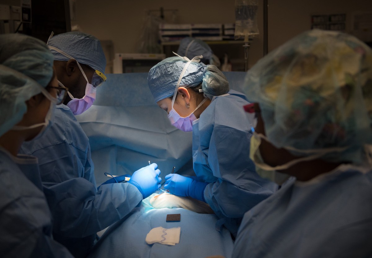
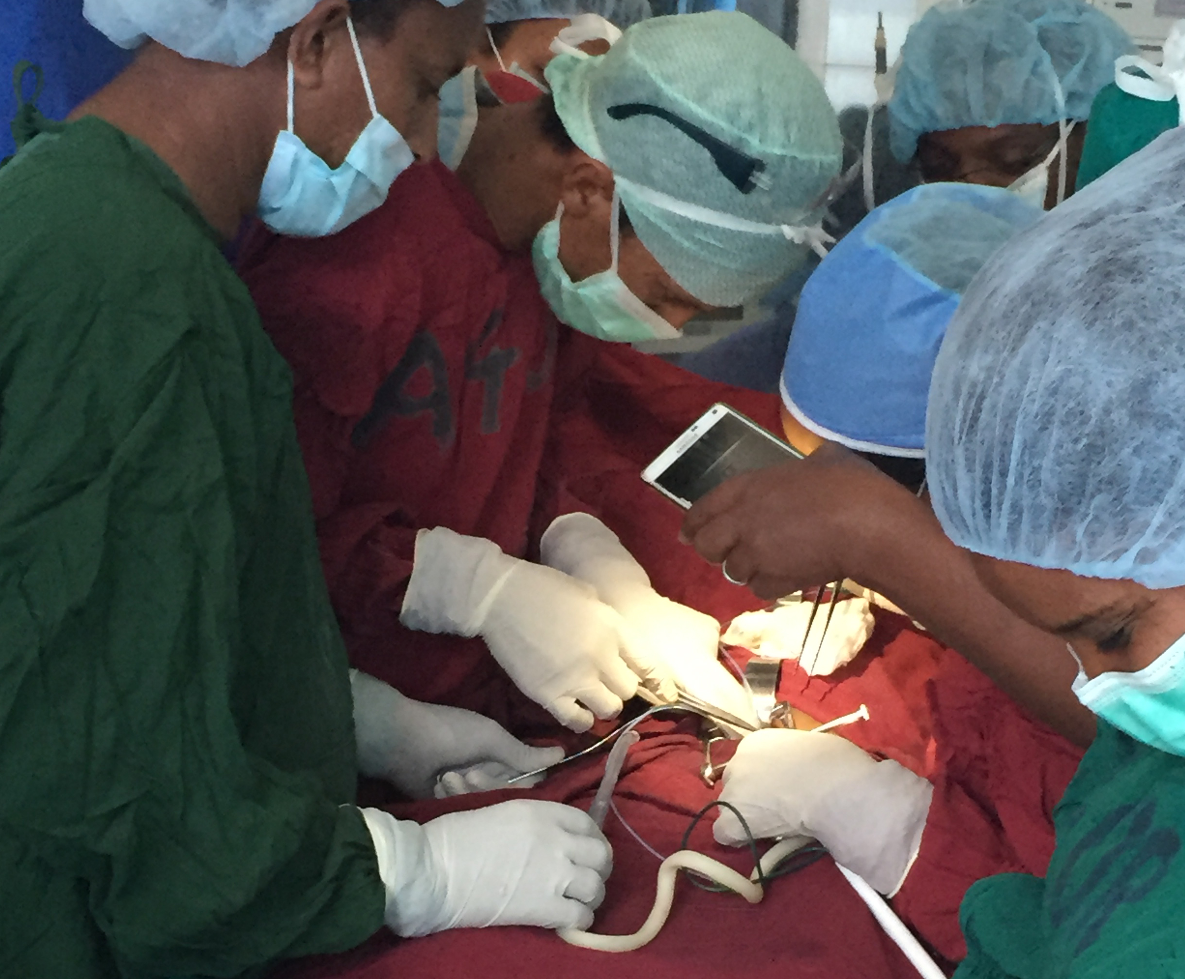
The Michigan team validated their technique with studies involving four participants with upper extremity amputations who had previously undergone RPNI surgery to treat neuroma pain. Each participant had a total of 3 to 9 muscle grafts implanted on nerves. Initially, the researchers measured the signals from these RPNIs using fine-wire, nickel-alloy electrodes, which were inserted through the skin into the grafts using ultrasound guidance. They measured high-amplitude electromyography signals, representing the electrical activity of the mini-muscles, when the participants imagined they were moving the fingers of their phantom hand. The ultrasound images showed the participants’ thoughts caused the associated specific mini-muscles to contract. These proof-of-concept measurements, however, were limited by the discomfort and movement of the percutaneous electrodes that pierced the skin.

Next, the team surgically implanted permanent electrodes into the RPNIs of two of the participants. They used a type of electrode commonly used for battery-powered diaphragm pacing systems, which electrically stimulate the diaphragm muscles and nerves of patients with chronic respiratory insufficiency to help regulate their breathing. These implanted electrodes allowed the researchers to measure even larger electrical signals—week after week from the same participant—by just plugging into the connector. After taking 5 to 15 minutes of calibration data, the electrical signals were translated into movement intent using machine learning algorithms and then passed on to a prosthetic hand. Both subjects were able to intuitively complete tasks like stacking physical blocks without any training—it worked on the first try just by thinking about it, says Chestek. Another key result is that the algorithm kept working even 300 days later.

“The ability to use the determined relationship between electrical activity and intended movement for a very long period of time has important practical consequences for the user of a prosthesis, because the last thing they want is to rely on a hand that is not reliable,” Clark says.

Although this clinical trial is ongoing, the Michigan team is now investigating how to replace the connector and computer card with an implantable device that communicates wirelessly, so patients can walk around in the real world. The researchers are also working to incorporate sensory feedback through the regenerative peripheral nerve interface. Their ultimate goal is for patients to feel like their prosthetic hand is alive, taking over the space in the brain where their natural hand used to be.

“People are excited because this is a novel approach that will provide high quality, intuitive, and very specific signals that can be used in a very straightforward, natural way to provide high degrees of dexterous control that are also very stable and last a long time,” Clark says.

Read the article in Science Translational Medicine.

Illustration of multiple regenerative peripheral nerve interfaces (RPNIs) created for each available nerve of an amputee. Fine-wire electrodes were embedded into his RPNI muscles during the readout session. Credit: Philip Vu/University of Michigan; Science Translational Medicine doi: 10.1126/scitranslmed.aay2857

This is a reposting of my news brief, courtesy of Materials Research Society.

Behind the scenes with a Stanford pediatric surgeon

In a new series, “Behind the Scenes,” we’re inviting Stanford Medicine physicians, nurses, researchers and staff to share a glimpse of their day.

As a science writer, I talk to a lot of health care providers about their work. But I’ve often wondered what it is really like to be a surgeon. So I was excited to speak with pediatric surgeon Stephanie Chao, MD, about her day.

Chao is a pediatric general surgeon, an assistant professor of surgery and the trauma medical director for Stanford Children’s Health. In addition to performing surgeries on children of all ages, she has a range of research interests, including how to reduce gun-related deaths in children and the hospital cost associated with pediatric firearm injuries.

Morning routine
On days that I operate, I get up between 5:50 and 6 a.m., depending on whether I hit the snooze button. I typically don’t eat breakfast. I don’t drink coffee because I don’t want to get a tremor. I’m out the door by 6:30 a.m. and at the hospital by 7 a.m. I usually go by the bedside of the first patient I’m going to operate on to say hi. The patient is in the operating room by 7:30 a.m. and my cases start.

On non-surgical days, it’s more chaotic. I have a 3-year-old and 1-year-old. So every day there’s a jigsaw puzzle as to whether my husband or I stay to get the kids ready for preschool, and who comes home early.

Part of Stephanie Chao’s day involves checking on patients, including this newborn.

In the operating room
The operating room is the place where I have the privilege of helping children feel better. It’s a very calming place, like a temple. When I walk through the operating room doors, the rest of the world becomes quiet. Even if it is a high-intensity case when the patient is very sick, I know there is a team of nurses, scrub techs and anesthesiologists used to working together in a well-orchestrated fashion. So even when the unexpected arises, we can focus on the patient with full confidence that we’ll find a solution.

There are occasions when babies are so sick that we need silence in the operating room. Everyone becomes hyper-attuned to all the beeps on the monitors. When patients are not as critically sick, I often play a Pandora station that I created called “Happy.” I started it with Pharrell Williams’ “Happy” and then Pandora pulled in other upbeat songs, including a bunch of Taylor Swift songs, so everyone thinks I’m a big Taylor Swift fan.

The OR staff call me by my first name. I believe that if everyone is relaxed and feels like they have an equal say in the procedure, we work better as a well-oiled machine for the benefit of the patient.

“The OR staff call me by my first name,” Stephanie Chao said.

Favorite task
Some of the most rewarding times of my day are when I sit down with patients and their families to hear their concerns, to reassure them and to help them understand what to expect — and hopefully to make a scary situation a little less so. As a parent, I realize just how hard it is to entrust one’s child completely in the hands of another. I also like to see patients in the hospital as they’re recovering.

Favorite time
The best part of the day is when I come home. When I open the door into the house, my kids recognize that sound and I hear their little footsteps as they run towards the door, shrieking with joy.

Evening ritual
When my husband and I get home, on nights I am not on call, I cook dinner in the middle of the chaos of hearing about the kids’ day. Hopefully, we “sit down” to eat by 6:20 or 6:30 p.m., and I mean that term loosely. It’s a circus, but eventually everyone is somewhat fed.

And then we do bath time and bedtime. There’s a daily negotiation with my three-year-old on how many books we read before bed. On school nights, she’s allowed three books but she tries to negotiate for 10.

Eventually, we get both kids down for the night. Then my husband and I clean up the mess of the day and try to have a coherent conversation with each other. But by then both of us are exhausted. I try to log on again to finish some work, read or review papers. I usually go to sleep around 11 p.m.

Managing it all
When I can carve out time to do relaxing things for myself, like go to the gym, that is great. But it’s rare and I remind myself that I am blessed with a job that I love and a wonderfully active family.

The result sometimes feels like chaos, but I don’t want to wish my life away waiting for my kids to get older and for life to get easier. Trying to live in the moment, and embracing it, is how I find balance.

Photos by Rachel Baker

This is a reposting of my Scope blog story, courtesy of Stanford School of Medicine.

Designing an inexpensive surgical headlight: A Q&A with a Stanford surgeon

2017_Ethiopia_Jared Forrester_Black Lion_Pediatric OR by cell phone_2_crop
Photo by Jared Forrester / © Lifebox 2017

For millions of people throughout the world, even the simplest surgeries can be risky due to challenging conditions like frequent power outages. In response, Stanford surgeon Thomas Weiser, MD, is part of a team from Lifebox working to develop a durable, affordable and high-quality surgical headlamp for use in low-resource settings. Lifebox is a nonprofit that aims to make surgery safer throughout the world.

Why is an inexpensive surgical headlight important?

“The least expensive headlight in the United States costs upwards of $1000, and most cost quite a bit more. They are very powerful and provide excellent light, but they’re not fit for purpose in lower-resource settings. They are Ferraris when what we need is a Tata – functional, but affordable.

Jared Forrester, MD, a third-year Stanford surgical resident, lived and worked in Ethiopia for the last two years. During that time, he noted that 80 percent of surgeons working in low- and middle-income countries identify poor lighting as a safety issue and nearly 20 percent report direct experience of poor-quality lighting leading to negative patient outcomes. So there is a massive need for a lighting solution.”

How did you develop your headlamp specifications?

“Jared started by passing around a number of off-the-shelf medical headlights with surgeons in Ethiopia. We also asked surgeons in the U.S. and the U.K. to try them out to see how they felt and evaluate what was good and bad about them.

We performed some illumination and identification tests using pieces of meat in a shoebox with a slit cut in it to mimic a limited field of view and a deep hole. We asked surgeons to use lights at various power with the room lights off, with just the room lights on and with overhead surgical lights focused on the field. That way we could evaluate the range of light needed in settings with highly variable lighting, something that does not really exist here in the U.S.”

How do they differ from recreational headlamps?

“Recreational headlights have their uses and I’ve seen them used for providing care — including surgery. However, they tend to be uncomfortable during long cases and not secure on the head. Also, the light isn’t uniformly bright. You can see this when you shine a recreational light on a wall: there is a halo and the center is a different brightness than the outer edge of the light. This makes distinguishing tissue planes and anatomy more difficult.”

What are the barriers to implementation?

“While surgeons working in these settings all express interest in having a quality headlight, there is no reliable manufacturer or distributor for them. Surgeons cannot afford expensive lights, and no one has stepped up to provide a low-cost alternative that is robust, high quality and durable. We’re working to change that.”

What are your next steps?

“We are now evaluating a select number of headlights and engaging manufacturers in discussions about their current devices and what changes might be needed to make a final light at a price point that would be affordable to clinicians and facilities in these settings. By working through our networks and using our logistical capacity, we can connect the manufacturer with a new market that currently does not exist  — but is ready and waiting to be developed.

We believe these lights will improve the ability of surgeons to provide better, safer surgical care and also allow emergency cases to be completed at night when power fluctuations are most problematic. These lights should increase the confidence of the surgeon that the operation can be performed safely.”

This is a reposting of my Scope blog story, courtesy of Stanford School of Medicine.

Space, the new surgical frontier? A Q&A

Photo by WikiImages

Many medical treatments — in their current form — would be unfeasible on deep space missions, such as a journey to Mars.

How will we diagnose and treat the ailments of future space travelers? And what medical issues will they likely encounter? I posed these questions to Sandip Panesar, MD, a postdoctoral research fellow at Stanford who wrote a recent article about surgery in space in the British Journal of Surgery.

What inspired you to research surgery in space?

“From a young age, I’ve always been interested in space travel. I also have a background in surgery, trauma and emergency medicine. So it just clicked one day when I was reading about SpaceX. I realized they may actually send people to Mars, so we need to consider the medical implications of that. Specifically, how would you perform surgery?

The need for surgical care in space in the near future will likely revolve around emergency situations — such as crushes, impacts, falls and burns — since the possibility of trauma occurring during exploratory missions can never be ruled out. In cases of severe trauma, significant internal bleeding may necessitate invasive surgical procedures.”

What adverse conditions do space travelers face?

“People are exposed to a few key physical conditions in space — solar particle radiation, temperature extremes and a lack of gravity. Solar particle radiation is a lot different than the particles people are exposed to on Earth. It has a higher chance of causing DNA damage, leading to an increased risk of high-grade cancers prone to metastasize. However, a lack of gravity causes a whole host of even more critical changes in the human body.”

How does this extraterrestrial environment impact human physiology?

“One of the biggest changes is the redistribution of bodily fluids. On Earth, gravity and walking upright pulls most of our fluids down to our legs. In space, these fluids distribute evenly throughout the body. This affects heart rate and blood pressure, increases intracranial pressure and causes face swelling. And it decreases leg size, a phenomenon called ‘chicken legs.’

An absence of gravity also causes the bones and muscles to atrophy.

In addition, the makeup of white blood cells changes in space. Plus, the body produces more stress hormones, called glucocorticoids, which further weaken the immune system. This may negatively affect wound healing, which is critical to surgical recovery.

Microbes are also known to be more pathological in space, making the risk of a serious infection after surgery even higher.”

How can surgery be adapted for space?

“One idea is to include a trauma pod, an enclosed medical suite, in the space station or vessel — a concept that originated in military medicine.

We’ve also proposed minimally-invasive keyhole surgery, but it has limited use in trauma situations and a pretty large learning curve. So open surgery is likely but challenging in space. For instance, the bowel is free-floating within the abdominal cavity,  so it can float out when you open the stomach if there’s no gravity. This carries a risk of infection, contamination and perforation. One potential solution is to use a hermetically sealed enclosure — placing a clear plastic box over the wound and working essentially in a glove box with a pressure differential.”

Could surgical robots or other equipment help?

“Mars is 48 million miles away and the radio signal delay is 20 minutes, so using robots controlled by surgeons on Earth isn’t feasible. Instead, researchers are developing robots that can perform surgery by themselves or with really minimal human assistance. There have already been trials of robots that can suture together pig bowels with minimal assistance.

Finally, the size and weight of the payload is a huge barrier and surgical specialties all use different tools. A feasible solution is to bring a 3D printer that can print bandages, casts, surgical tools and even maybe pharmaceuticals. Also, you could diagnosis with an ultrasound scanner and a compact CT scanner like the ones used in ambulances in the UK.”

Would you want to be an on-board surgeon?

“Not just yet. I still have a lot of things I want to do on Earth.”

This is a reposting of my Scope blog story, courtesy of Stanford School of Medicine.

New course highlights how surgeons can serve their communities

Photo courtesy of Jecca Steinberg

Stanford medical students Jecca Steinberg and Paloma Marin-Nevarez want to spread the word that service-minded medical students can care for underserved communities by specializing in surgery. With the help of their mentor James Lau, MD, they have created an upcoming seminar series for medical students called “Service Through Surgery,” which showcases how surgeons can address health inequities.

Beginning in January, the new 10-week course will expose Stanford medical students to a diverse group of surgical leaders who are passionate about improving health equity through surgery. I connected with Steinberg, shown on the left in the photo, and Marin-Nevarez to learn more.

What inspired you to create the Service through Surgery seminar course?

Marin-Nevarez: “I emigrated from Mexico when I was 10 and settled in a low-income community in south Los Angeles. I never really considered myself disadvantaged until I went to college and experienced firsthand the shortcomings of my education system. Ever since, I knew I would make my life’s work to serve the underserved in communities like my own.

In my second year of medical school, I fell in love with surgery. However, when I thought about being a ‘community physician,’ I didn’t see how surgery would fit into that picture. The speakers in this course will show students with the same internal struggle as mine that they don’t need to compromise their values in order to pursue their dreams.”

What role can diversity play in overcoming health inequities?

Steinberg: “Low-income, minority communities continue to receive inadequate surgical services and bear unconscionable health burdens. Research has demonstrated that increasing diversity among physicians improves healthcare access and outcomes for traditionally disenfranchised communities, but surgery continues to trail behind other medical specialties in racial, socioeconomic and gender diversity. So the surgical workforce represents an underutilized resource for decreasing health inequities and improving the health of our communities.”

Marin-Nevarez: “A more diverse workforce leads to better outcomes for the underserved because minority patients are more likely to seek care from and be more comfortable with physicians from diverse backgrounds. And physicians from diverse backgrounds are more likely to treat patients of color in underserved communities.”

What causes surgery to be less diverse than other medical specialties?

Marin-Nevarez: “Because of unequal opportunities — especially for communities of color — surgeons are not as diverse as they should be. Because of this lack of diversity, there is a lack of mentorship that then perpetuates the cycle.

Mentorship can make a huge difference in recruiting people into a field. For example, James Lau, MD, is an amazing mentor — he was the first person to make me believe that being the first surgeon in my family may be an attainable goal. Those who ‘make it’ without mentorship most likely had access to extra resources or had to work much harder than their counterparts, or both.”

How will your seminar course inspire change?

Steinberg: “Our seminar course will create an opportunity for Stanford medical students to meet and form relationships with accomplished physicians who have combined their passions for diminishing inequities and surgery. It will show the incredible impact surgeons can make on their community. For example, Matias Bruzoni, MD, will talk about a Spanish clinic he created from scratch to improve the surgical experiences and outcomes of Spanish speaking patients. And Sherry Wren, MD, will provide her perspective on serving veterans domestically and populations around the world, exploring the adversity she faced in dedicating her career to social service.

When students connect with role models like these with a similar background and passions, they are more likely to follow in the trajectory of that role model and consider careers that might have previously seemed unattainable. We hope this seminar will provide that initial connection.”

This is a reposting of my Scope blog story, courtesy of Stanford School of Medicine.

Stanford researchers develop simulations to improve heart surgeries

MRI or CT scans provide physicians with a detailed picture of their patients’ internal anatomy. Heart surgeons often use these images to plan surgeries.

Unfortunately, these anatomical images don’t show how the blood is flowing through the vessels — which is critical, according to Alison Marsden, PhD, a Stanford associate professor of pediatrics and of bioengineering. In the video above, she explains that many surgeons currently use a pencil and paper to sketch out their surgical plan based on the patient’s images. She hopes to change this.

Marsden and her colleagues at Stanford’s Cardiovascular Biomechanics Computational Lab are developing a new technique — using imaging data and specialized simulation software — to predict what is likely to happen during heart surgery.

“What we’re trying to do is bring in that missing piece of what are these detailed blood flow patterns and what might happen if we go in and make an intervention, for example, opening up a blocked blood vessel or putting in a bypass graft,” Marsden said in a recent Stanford Engineering news story.

Their open source software, called SimVascular, loads the imaging data, constructs a 3D anatomical model of the heart and then simulates the patient’s blood flow. It has already been used to help design the surgical plan for several babies born with a severe form of congenital heart disease, Marsden said. However, more research is needed to determine whether the technique improves patient outcomes before it can be widely used in the clinic.

This is a reposting of my Scope blog story, courtesy of Stanford School of Medicine.

From art to surgery: Stanford alumna reconstructs new ears for children

Dr Sheryl Lewin in the operating room (courtesy of Lewin)

Some children are born with a missing or malformed small ear due to a rare congenital condition called microtia. In most cases, the child’s ear canal is also very small or absent, resulting in hearing loss.

The surgical procedures used to correct microtia require the skills of both a sculptor and surgeon — making it the perfect specialty for Sheryl Lewin, MD, a craniofacial plastic surgeon who began her training as an artist and architect.

Lewin’s career has been passionately devoted to treating microtia through her private medical practice and nonprofit organization called Earicles, which helps children born without ears through education, research and free or reduced-cost treatment. I recently spoke with Lewin about her work: 

As an architect major, what inspired you to become a physician?

“When I was in architecture school at UC Berkeley, I loved the challenge of design, where you can use your own creativity to solve visual and spatial problems. My program was heavily artistic — we drew, painted and sculpted. But what was missing was the ability to use those skills to directly affect someone’s life in a tangible and meaningful way.

During college, I lived across the street from an elementary school that served underprivileged kids, which inspired me to start a volunteer organization of Berkeley undergraduates that mentored disadvantaged children in the local community. I recognized that I really enjoyed working and helping kids, and medicine was a way to do that.

When I went to medical school at Stanford, I was drawn to surgery as it gave me the ability to work with my hands. I decided to pursue pediatric plastic surgery after I saw my first cleft lip surgery on a tiny infant, whose life was transformed in a couple of hours. I realized it absolutely used the same skill set that I was used to working in: design, thinking three-dimensionally and visualizing symmetry. It was very much like sculpture.

Years later in medical school, I saw my first surgery to correct a rare condition called microtia. Once I saw what was involved, there was no doubt that I would love the challenge of making ears, which is considered by many plastic surgeons to be one of the most technically difficult things we do. But what really sealed the deal was the intangible feeling you get taking care of these children and their families. I came home that day and told my husband, ‘I know what I want to do with the rest of my life.’”

What is microtia?

Microtia ear before and after surgery (Courtesy of Lewin)
Child’s ear before and after microtia reconstruction surgery (courtesy of Lewin)

“Microtia is a congenital condition in which the ear does not develop properly. The word microtia translates to “small ear.” It occurs in about one in 6,000 to 12,000 children worldwide, with a higher prevalence among Hispanics, Asians and Native Americans.

The cause of microtia is not well understood, particularly regarding the role of environmental and genetic factors. Some medications have been linked to microtia when ingested in the first trimester of pregnancy, including Thalidomide and Accutane. However, it’s important to understand that microtia is rarely caused by what a mother does during pregnancy.”

How do you treat microtia? 

“Ninety-five percent of the world treats microtia by removing rib cartilage from the chest, carving it into an ear framework and then slipping it under the skin. In order to have enough cartilage, surgery must be delayed until children are six to ten years old. Three to four surgeries are required with this technique, and the ability to match the opposite ear is limited.

Several colleagues and I use a different technique. In an eight to ten hour outpatient surgery, I customize a porous polyethylene implant into a three-dimensional ear shape that matches the opposite ear. This biocompatible implant is then covered with vascular tissue. This allows for a symmetric and natural appearing ear to be created in just one operation as early as three years of age.

Children with microtia almost always have conductive hearing loss — since the ear canal is missing but the auditory nerve is functional. During the ear reconstruction surgery, I can do an additional scarless procedure to help restore hearing. I implant a titanium device in the skull that is connected to a bone conduction hearing processor, commonly referred to as a BAHA. The hearing processor captures sound and transmits these vibrations to the skull through the implant, which stimulates the auditory nerve that processes it as sound.”

What is most rewarding about your work?

“This surgery not only helps provide functionality, such as giving children the ability to wear eyeglasses, but it’s often about helping children attain the simplest human need: to feel the same as everyone else.

One of my favorite moments involved a four-year old boy named Davin, who had microtia of both ears. I was sitting next to him as he was about to see his second ear for the first time. He looked me in the eyes and said, “Dr. Lewin, do I have two big boy ears now?” I said, “Yes Davin, two beautiful ears.” Then, out of nowhere, he leaned over and planted a big kiss right on my lips and said, “Dr. Lewin, I love you.” For a moment, I was speechless, and then managed to say, “Davin, I love you too.” It really can’t get any better than that in my book.”

This is a reposting of my Scope blog story, courtesy of Stanford School of Medicine.

Surgery to find your voice: A Q&A with expert Anna Messner

5540462170_5ca4645115_o_flickr_HowardLake_560x.315jpg
Photo by Howard Lake

When we’re in a noisy restaurant, it’s really difficult to hear my young niece speak. She can only talk very quietly, because she has a paralyzed vocal cord.

Like many children born very premature, the nerve going to her vocal cord was likely damaged when she had heart surgery soon after she was born. Her inability to be heard frustrates her, especially now that she is in school. However, a rare surgery may bring her the hope of a near-normal voice.

Stanford surgeons recently began performing laryngeal reinnervation surgery, which essentially rewires the paralyzed vocal cord with a new nerve supply. I recently spoke with Anna Messner, MD, a professor of otolaryngology and pediatrics who sees patients at Lucile Packard Children’s Hospital Stanford, about laryngeal reinnervation surgery.

What standard surgical procedures are used to treat unilateral vocal cord paralysis?

In general, the surgical procedures bulk up the paralyzed vocal cord to move it towards the midline of the body, making it easier for the other vocal cord to compensate and close. There are two standard surgeries. We can do injection laryngoplasty, where we inject a substance into the paralyzed vocal cord to thicken it. Unfortunately, this procedure often needs to be repeated multiple times, if it works at all. We can also insert a medialization implant in teenagers and adults, but this doesn’t work for growing kids. If we put an implant into a two year old, it wouldn’t be an appropriate size when he is 10.

How does laryngeal reinnervation surgery work?

No matter what we do, we can’t make the vocal cord move. We can never make it perfect again. What we can do is hook up one of the other nerves in the neck to the recurrent laryngeal nerve that goes to the vocal cord. And that helps some new nerve fibers go to the vocal cord, making the vocal cord stronger and thicker. As a result, the voices on these kids improve significantly.

The surgery itself is fairly straightforward and only takes about an hour. The children typical go home the same day or just stay overnight, and they feel back to normal in a couple of days. But then we have to wait five to six months for the nerve fibers to grow before we can see real improvement in the voice. The only downside is that it takes a long time to see the effects of the surgery.

What inspired you to learn the laryngeal reinnervation procedure?

We have a large pediatric cardiac surgery program at Stanford, so we have quite a few patients with vocal cord paralysis. Most of our patients are born prematurely and need heart surgery, which can pull and damage the nerve that goes to the vocal cord on one side. After these surgeries, the damaged vocal cord starts working again in just over a third of the cases. But for the rest of the kids, the vocal cord remains paralyzed.

The standard surgeries just don’t work very well, so we’ve had a longstanding interest in finding alternatives. I saw Marshall Smith, MD, the medical director of the Voice Disorders Center at University of Utah, give a presentation on his clinical trials. So I observed him performing the reinnervation surgery about 1.5 years ago, and since then I’ve been performing the surgery. One of my colleagues, Doug Sidell, MD, also performs the surgery.

How does the voice improvement impact the patients?

The voice improvement makes a huge impact on the children, especially in school. For instance, when the children are trying to read a story or give a presentation in front of the classroom, now they can actually be heard. The results are very encouraging. The surgery has the potential for huge, life-long voice improvement.

This is a reposting of my Scope medical blog story, courtesy of Stanford School of Medicine.

New Approach to Using Stem Cells During Orthopedic Surgery

Device used by UC Davis researchers to rapidly concentrate stem cells, which are harvested from surgical irrigation fluid during an orthopedic procedure (Courtesy of SynGen Inc).

About 6 million people in North America suffer bone fractures each year and 5 to 10 percent of these patients are resistant to healing, according to the American Academy of Orthopaedic Surgeons. This means that about half a million Americans annually have fractures that don’t heal. UC Davis researchers are developing an improved surgical therapy for such fractures, using stem cells and innovative technology.

After a broken bone is treated, new bone tissue usually begins to form and connect the broken pieces. However, some bone fractures don’t heal due to a lack of adequate stability, blood flow, or large bone loss. For instance, severe bone fractures that are caused by a high-energy car wreck are more likely not to heal. Several other factors increase the risk of non-healing bones, including older age, diabetes, poor nutrition, use of tobacco, and severe anemia. Traditional treatments to address this problem, such as bone grafts taken from another part of the body, often lead to pain, dysfunctional limbs, and disabilities.

In the last several years, the application of stem cells directly to the wound site has emerged as an improved way to treat non-healing fractures. However, acquiring the necessary stem cells from the patient, a matched donor, or embryo is problematic. Ideally the stem cells come directly from the patient, but this requires a painful surgical procedure with general anesthesia during which a large needle is used to retrieve the stem cells from the hip. In addition, the retrieved stem cells need to be isolated before they can be transplanted back into a patient, so a second surgery is required with a long combined recovery period.

“People come to me after suffering for six months or more with a non-healing bone fracture, often after multiple surgeries, infections and hospitalizations,” said Mark Lee, UC Davis associate professor of orthopaedic surgery, in a press release. “Stem cell therapy for these patients can be miraculous, and it is exciting to explore an important new way to improve on its delivery.”

Mark Lee, UC Davis associate professor of orthopedic surgery (Courtesy of UC Davis).

In their new clinical trial, Lee’s team is testing a new SynGen Inc. device that processes the irrigation fluid obtained during an orthopedic procedure. This irrigation fluid contains abundant mesenchymal stem cells and other factors that can be used to help make new blood vessels and improve wound healing.

During the surgery, the irrigation fluid is aspirated and captured. The fluid is then centrifuged and processed using the new SynGen device, which rapidly isolates a concentration of mesenchymal stem cells in less then 30 minutes. These concentrated stem cells are then delivered to the patient’s fracture during the same surgery. The device is about the size of a food processor, so it can be easily used in an operating room.

“The device’s small size and rapid capabilities allow autologous stem cell transplantation to take place during a single operation in the operating room rather than requiring two procedures over a period of weeks,” said Lee in the press release. “This is a dramatic difference that promises to make a real impact on wound healing and patient recovery.”

The UC Davis researchers are already testing this new surgical treatment on patients. However, it is unclear when this treatment could move into general clinical practice.

A modified version of this story is posted on my KQED Science blog.

%d bloggers like this: