Stanford researchers grow neural stem cells more efficiently in 3-D gel

Photo by luismmolina/Getty Images

Stem cells have the potential to help us understand and treat a range of diseases and injuries — from vision loss to cancers. For instance, a Japanese man in his 60s was recently treated for vision loss due to macular degeneration using stem cells donated by another person. And many other clinical trials involving stem cells are underway.

However, there is still a lot to learn about stem cells and many barriers to overcome before most potential treatments can be realized. One such barrier is how to grow large quantities of stem cells while maintaining their unique properties. Now, Stanford researchers have developed a new gel in which they can grow massive numbers of neural stem cells in less space.

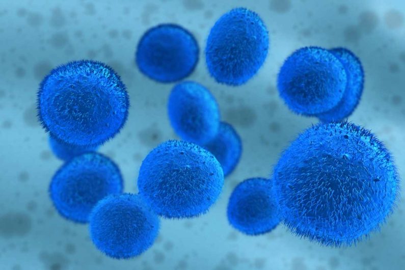
Stem cells are unspecialized cells that can self-renew and develop into many different types of cells in the body. Researchers hope that neural stem cells — that differentiate into neurons and glia cells in the nervous system — can be used to treat spinal cord injuries, Parkinson disease, Huntington disease and other nervous system disorders.

As recently reported in Nature Materials, the Stanford team engineered a new polymer-based gel optimized for neural stem cells, growing them in three dimensions instead of two.

“For a 3-D culture, we need only a 4-inch-by-4-inch plot of lab space, or about 16 square inches. A 2-D culture requires a plot of four feet by four feet, or about 16 square feet,” said the study’s first author Chris Madl, PhD, a postdoctoral research fellow in microbiology and immunology at Stanford, in a recent Stanford news release. In addition to taking 100-times less lab space, the new 3-D process also demands less energy and nutrients to grow the cells, he said.

A key to the development was the realization that neural stem cells need to chemically or physically remodel their surrounding environment to maintain their ability to differentiate into other cells. The researchers discovered this by creating and testing a family of gels with varying stiffness and remodeling susceptibilities. The authors explained in the paper, “Whereas cells cultured in 2-D are unrestricted and free to spread, cells within nanoporous 3-D hydrogels require matrix remodeling to spread, migrate, and proliferate.”

Surprisingly, they also discovered that the neural stem cells weren’t sensitive to the stiffness of the gel, unlike most other stem cells.

These new findings have given the leader of the research group new hope for future stem cell therapies. Sarah Heilshorn, PhD, associate professor of materials science and engineering at Stanford, said in the release, “There’s this convergence of biological knowledge and engineering principles in stem cell research that has me hopeful we might finally actually solve big problems.”

This is a reposting of my Scope blog story, courtesy of Stanford School of Medicine.

Stanford study provides new understanding of breast growth disorders

Photo by sasint

Breast underdevelopment at puberty is associated with a shortage of several hormones produced by the pituitary gland, a condition called combined pituitary hormone deficiency (CPHD). This disorder is caused in part by loss-of-function mutations of the GLI2 gene, but the molecular pathways of how CPHD manifests are not fully understood.

Now, researchers at Stanford University School of Medicine have discovered a new way that GLI2 impacts breast development, as recently reported in Science. Led by Philip Beachy, PhD, a Stanford professor of developmental biology and of biochemistry, the research team found that GLI2 activity helps control mammary stem cells in mice.

Stem cells are responsible for the growth, homeostasis and repair of many tissues. The behavior and survival of these stem cells depends on their local microenvironment, called a stem cell niche. During breast growth, the niche must support its associated stem cells while also responding to circulating hormones that trigger the dramatic changes of puberty.

The study showed that this stem cell niche is genetically programmed to produce the signals that control breast development in response to the hormones that regulate puberty. Using mice without a functioning GLI2 gene, the researchers found that a defective stem cell niche environment may lead to the breast growth defects seen in human CPHD. In addition, the research provides insights into a new mechanism to target when developing drugs that may help prevent breast cancer.

The authors conclude:

“Whereas prior studies implicate stem cell defects in human disease, this work shows that niche dysfunction may also cause disease, with possible relevance for human disorders and in particular the breast growth pathogenesis associated with combined pituitary hormone deficiency.”

This is a reposting of my Scope blog story, courtesy of Stanford School of Medicine.


New Approach to Using Stem Cells During Orthopedic Surgery

Device used by UC Davis researchers to rapidly concentrate stem cells, which are harvested from surgical irrigation fluid during an orthopedic procedure (Courtesy of SynGen Inc).

About 6 million people in North America suffer bone fractures each year and 5 to 10 percent of these patients are resistant to healing, according to the American Academy of Orthopaedic Surgeons. This means that about half a million Americans annually have fractures that don’t heal. UC Davis researchers are developing an improved surgical therapy for such fractures, using stem cells and innovative technology.

After a broken bone is treated, new bone tissue usually begins to form and connect the broken pieces. However, some bone fractures don’t heal due to a lack of adequate stability, blood flow, or large bone loss. For instance, severe bone fractures that are caused by a high-energy car wreck are more likely not to heal. Several other factors increase the risk of non-healing bones, including older age, diabetes, poor nutrition, use of tobacco, and severe anemia. Traditional treatments to address this problem, such as bone grafts taken from another part of the body, often lead to pain, dysfunctional limbs, and disabilities.

In the last several years, the application of stem cells directly to the wound site has emerged as an improved way to treat non-healing fractures. However, acquiring the necessary stem cells from the patient, a matched donor, or embryo is problematic. Ideally the stem cells come directly from the patient, but this requires a painful surgical procedure with general anesthesia during which a large needle is used to retrieve the stem cells from the hip. In addition, the retrieved stem cells need to be isolated before they can be transplanted back into a patient, so a second surgery is required with a long combined recovery period.

“People come to me after suffering for six months or more with a non-healing bone fracture, often after multiple surgeries, infections and hospitalizations,” said Mark Lee, UC Davis associate professor of orthopaedic surgery, in a press release. “Stem cell therapy for these patients can be miraculous, and it is exciting to explore an important new way to improve on its delivery.”

Mark Lee, UC Davis associate professor of orthopedic surgery (Courtesy of UC Davis).

In their new clinical trial, Lee’s team is testing a new SynGen Inc. device that processes the irrigation fluid obtained during an orthopedic procedure. This irrigation fluid contains abundant mesenchymal stem cells and other factors that can be used to help make new blood vessels and improve wound healing.

During the surgery, the irrigation fluid is aspirated and captured. The fluid is then centrifuged and processed using the new SynGen device, which rapidly isolates a concentration of mesenchymal stem cells in less then 30 minutes. These concentrated stem cells are then delivered to the patient’s fracture during the same surgery. The device is about the size of a food processor, so it can be easily used in an operating room.

“The device’s small size and rapid capabilities allow autologous stem cell transplantation to take place during a single operation in the operating room rather than requiring two procedures over a period of weeks,” said Lee in the press release. “This is a dramatic difference that promises to make a real impact on wound healing and patient recovery.”

The UC Davis researchers are already testing this new surgical treatment on patients. However, it is unclear when this treatment could move into general clinical practice.

A modified version of this story is posted on my KQED Science blog.

%d bloggers like this: