The number of cancer patients receiving proton beam therapy (PBT) – a newer, more targeted form of radiation therapy – is increasing, but Black patients are less likely to get this treatment than white patients, according to two recent studies published in JAMA Network Open.
Computers have revolutionized many fields, so it isn’t
surprising that they may be transforming cancer research. Computers are now
being used to model the molecular and cellular changes associated with individual
tumors, allowing scientists to simulate the tumor’s response to different combinations
of chemotherapy drugs.
Modeling big data to improve personalized cancer treatment
was the focus of a recent
episode of the Sirius radio show “The Future of Everything.” On hand was Sylvia Plevritis, PhD,
a professor of biomedical data science and of radiology at Stanford, who discussed
her work with Stanford professor and radio show host Russ Altman, MD, PhD.
Plevritis and her colleagues are using multi-omics
data — including measures of gene expression, protein function, metabolic
processes and more — to extensively profile individual tumors of individual
patients.
They are analyzing this data to better understand how tumors
become drug-resistant. She explained in the podcast that tumors are often heterogeneous
— not every cell has the same gene mutations — but chemotherapy drugs typically
target specific genetic mutations. Tumors are also driven by complex mechanisms
beyond genetic mutations. So her lab is comprehensively characterizing the
different cell types in a tumor and how these different cell types respond to
individual drugs. By better understanding the complexity of what drives the
tumor’s response, they hope to identify the underlying mechanisms of drug
resistance.
The goal, Plevritis said, is to more accurately estimate the
response of the entire tumor to a given set of drugs without having to run
clinical trials on every drug combination. Using their modeling, they hope to identify
the most promising drug combinations to make clinical trials more efficient, she
said.
The research team tested their computational model by
measuring the multi-omics profile of human cancer cells in a dish, before and
after exposing the cells to specific drugs. Their model then identified the
minimum combination of drugs with the maximum effect. This work used archived
cell samples, so their modeling results didn’t impact the patients’ treatment.
But they compared their model’s prediction to what drugs the patients actually received.
They determined that the best chemotherapy cocktail for most
of the patients would have been just one or two of the drugs that they
received. For about 10 percent of the patients, they predicted that a totally
different drug would have been the most effective, Plevaritis said in the
podcast.
Thus, their computational model may be able to divide
patients into different groups, based on tumor characteristics, and match those
groups with specific chemotherapy cocktails that would be most effective for
them. Plevaritis’ team is currently setting up a study to validate their computational
predictions for a group of patients with acute myeloid leukemia, in parallel
with a combination drug therapy trial, she said.
As a member of the Cancer Intervention Surveillance Network Modeling consortium, Plevritis is also using computational models to evaluate the impact of cancer screening guidelines — such as the recommended frequency of mammograms for general breast cancer screening — on mortality rates. For example, policy organizations like the U.S. Preventive Service Task Force often ask the consortium to simulate thousands of different screening policies — and rank their potential impact — to use as part of their selection criteria, she said.
One outcome of this work is an online decision tool for women who are
at high risk for developing breast cancer because they carry a mutation in the
BRCA1 or BRCA2 gene. Plevritis said about 45,000 people worldwide have used the
tool, and her team has received a lot of positive feedback.
“It’s been very satisfying to get these emails and this feedback from individuals who feel that this complex information was distilled in a way that they can make sense of it,” Plevritis said.
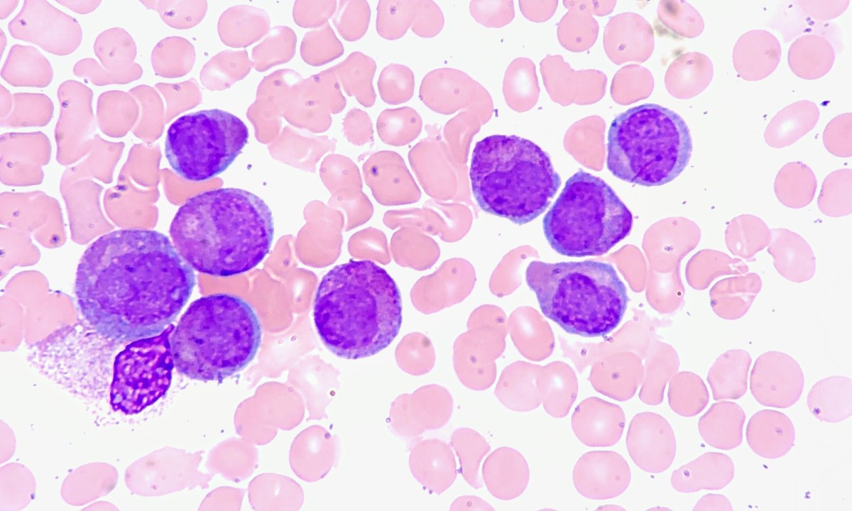
Image of acute promyelocytic leukemia cells by Ed Uthman
This is a reposting of my Scope blog story, courtesy of Stanford School of Medicine.
As a cancer survivor, I know how finishing treatment can
feel both happy and unsettling. I was ecstatic to be done with chemotherapy and
radiation therapy, but I worried about recurrence and long-term treatment side
effects. Whenever I went to a cancer checkup, I wore silly socks to remind
myself to smile. And decades later, I still have to be vigilant with periodic screening
tests like breast MRIs due to increased health risks from radiation.
Quality survivorship care requires a strong collaboration
between oncology and primary care clinicians, particularly as patients complete
their treatment. To help patients during this critical time, Stanford is
piloting a cancer survivorship clinic embedded in the practice of primary care
physician Jennifer Kim, MD. I
recently corresponded with her to learn more.
What inspired you to focus on cancer
survivorship care?
“When medical oncologist Lidia Schapira, MD, and I met about 2 years ago, we discussed her ideas of integrating primary care into survivorship at Stanford. All primary care practices, including my own, already care for survivors. As this is not a topic that I learned about in training, I wasn’t sure what would be different about calling it survivorship.
I learned more by attending national conferences and reviewing online curricula on survivorship. I also spent clinic days with oncologists as they saw patients. As I started to learn about survivorship, I realized that cancer and cancer treatment changes all aspects of patients’ health — medical, emotional and social — for the rest of their lives.”
How does your clinic work?
“Together with Lidia Schapira, I started Stanford’s Primary Care Cancer Survivorship clinic. I’m currently the only primary care physician doing this at Stanford and I see patients for two half days per week in the Hoover primary care clinic. My visits are consultative, meaning patients come to see me for one to several visits to discuss a complete survivorship plan, which they can bring back to their primary care physician for ongoing care.
The focus of
my visits is to detail a full treatment history and make a personalized
survivorship plan for issues such as cancer surveillance, potential long-term and
late effects of treatment, psychosocial concerns, co-morbidities and preventative care. I create this
history and plan together with the patient, so both the patient and their whole
care team will understand the content.
Having the clinic embedded in my primary care clinic — a different building and environment than the oncology department — helps us physically and mentally shift gears and transition to a primary care-based survivorship plan.”
What have you learned?
“I’ve learned the most powerful survivorship lessons from my patients and their experiences. I’ve learned not to assume what my patients are struggling with. Instead, by asking about their experiences and listening to their concerns, I can better understand what is really important to each individual. I’ve also found that it is very important to be open about and sensitive to emotional and psychosocial issues, including fear of recurrence, anxiety, fertility and sexual health. These topics are rarely the focus of oncology visits and patients don’t know who to ask.
I now realize that survivors often struggle with the transition from oncology care back to primary care-based care. It’s a challenging, overwhelming and emotional time when many still have significant long-term effects of treatment and multiple specialist visits. Patients often voice a need for a ‘quarterback’ to help guide them through the next phase of recovery — finding health after cancer.”
Do you have any advice for other
primary care physicians?
“Primary care physicians can and should be an essential part of survivorship and health after cancer. However, there are currently many barriers to survivorship being integrated into primary care — a knowledge gap, disparate electronic medical records, limited appointment time and patient concerns over whether primary care physicians are able to manage survivorship.
Many primary care physicians aren’t confident in their own survivorship knowledge, as there are so many cancers and so many treatments to keep track of, even in terms of surveillance recommendations and potential long-term effects. This is why shared care with specialists and continuing education can make a great impact in this area of increasing need.
With the help
of a great team, Lidia and I are developing an online course with video,
animation and text to help primary care physicians gain more knowledge, resources
and confidence in their long-term care of survivors. We hope to distribute this
widely when it is ready.
We’ve also started to create a patient-facing survivorship course that will focus on self-management, communication and resources. We hope this will help patients better navigate survivorship issues on their own and with their care team.”
Photo by Pamela Williams
This is a reposting of my Scope blog story, courtesy of Stanford School of Medicine.
The guidelines
for screening women for breast cancer are a bit confusing. The American Cancer Society
recommends annual mammograms for women older than 45 years with average risk, but
other groups like the U.S. Preventative Services Task Force (USPSTF) recommend
less aggressive breast screening.
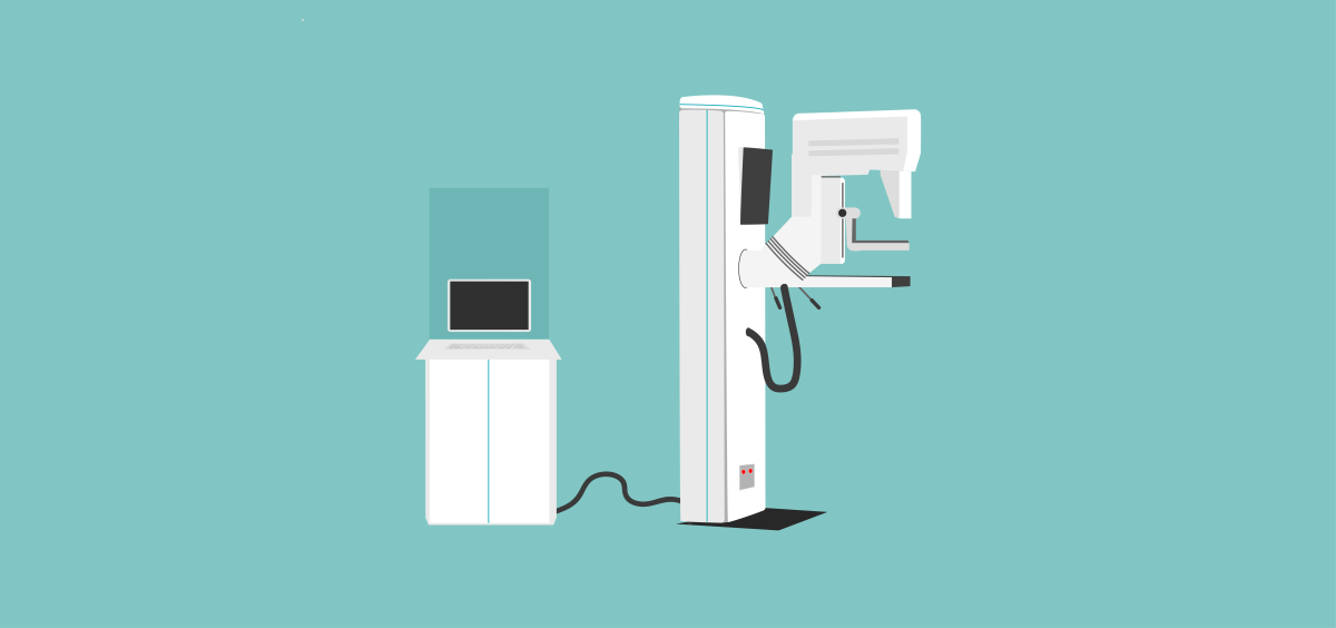
This controversy centers on mammography’s frequent false-positive
detections — or false alarms — which lead to unnecessary stress, additional
imaging exams and biopsies. USPSTF argues that the harms
of early and frequent mammography outweigh the benefits.
However, a recent Stanford study
suggests a better way to reduce these false alarms without increasing the
number of missed cancers. Using over 112,000 mammography cases collected from
13 radiologists across two teaching hospitals, the researchers developed and tested
a machine-learning model that could help radiologists improve their mammography
practice.
Each mammography case included the radiologist’s
observations and diagnostic classification from the mammogram, the patient’s
risk factors and the “ground-truth” of whether or not the patient had breast cancer
based on follow-up procedures. The researchers used the data to train and evaluate
their computer model.
They compared the radiologists’ performance against their machine-learning model, doing a separate analysis for each of the 13 radiologists. They found significant variability among radiologists.
Based on accepted clinical
guidelines, radiologists should recommend follow-up imaging or a biopsy when
a mammographic finding has a two percent probability of being malignant. However,
the Stanford study found participating radiologists used a threshold that
varied from 0.6 to 3.0%. In the future, similar quantitative observations could
be used to identify sources of variability and to improve radiologist training,
the paper said.
The study included 1,214 malignant cases, which represents
1.1 percent of the total number. Overall, the radiologists reported 176 false
negatives indicating cancers missed at the time of the mammograms. They also
reported 12,476 false positives or false alarms. In comparison, the
machine-learning model missed one additional cancer but it decreased the number
of false alarms by 3,612 cases relative to the radiologists’ assessment.
The study concluded: “Our results show that we can
significantly reduce screening mammography false positives with a minimal
increase in false negatives.”
However, their computer model was developed using data from
1999 to 2010, the era of analog film mammography. In future work, the
researchers plan to update the computer algorithm to use the newer descriptors
and classifications for digital
mammography and three-dimensional breast
tomosynthesis.
Ross Shachter, PhD, a Stanford associate professor of management science and engineering and lead author on the paper, summarized in a recent Stanford Engineering news release, “Our approach demonstrates the potential to help all radiologists, even experts, perform better.”
This is a reposting of my Scope blog story, courtesy of Stanford School of Medicine.
Neurobiologist and activist Martin Inderbitzen, PhD, began
his talk with a question: “Did you ever face a life situation that was totally
overwhelming?” Most of his audience likely answered yes, since he was speaking
to cancer survivors and their families at a Stanford event called Celebrating
Cancer Survivors.
The evening focused on life after cancer and highlighted Stanford’s
Cancer
Survivorship Program, which helps survivors and their families transition to
life after treatment by providing multidisciplinary services and health care. Lidia Schapira, MD, a
medical oncologist and director of the program, said they aim to “help people back into health.”
But to me, the heart of the event was the personal stories openly
shared by the attendees while standing in line for the food buffet or waiting
for the speeches to begin. As a Hodgkin’s survivor who was treated at Stanford twenty-five
years ago, I swapped “cancer stories” with my comrades.
Inderbitzen understands firsthand the importance of sharing such
cancer survival stories. In 2012, he was diagnosed at the age of 32 with pancreatic
cancer. From an online search, he quickly learned that 95 percent of people
with his type of cancer die within a few years. However, his doctor gave him
hope by mentioning a similar patient, who was successfully treated some years
earlier and is now happily skiing in the mountains.
“This picture of someone skiing in the mountains became my
mantra,” Inderbitzen explained. “I had all these bad statistics against me, but
then I also had this one story. And I thought, maybe I can also be one story,
because this story was somehow the personification of a possibility. It
inspired me to rethink how I saw my own situation.”
Later, Inderbitzen publicly shared his own cancer journey,
which touched many people who reached out to him. This inspired him to found MySurvivalStory.org — an initiative
that documents inspiring cancer survival stories to help other cancer patients better
cope with their illness. He and his wife quit their jobs, raised some funds and
began traveling around the globe to find and record short videos of cancer
survivors from different cultures.
“We share the stories in formats that people can consume
when they have ‘chemo brain’ — like podcasts you can listen to and short videos
you can process even when you’re tired,” he said. He added, “These stories are
powerful because they provide us with something or someone to aspire to —
someone who is a bit ahead of us, so we think “I can do that.’”
Inderbitzen isn’t the only one to recognize the empowering impact
of telling your cancer story. For example, the Stanford Center for Integrative
Medicine compiles some patient
stories on their Surviving Cancer website. And all of these stories have
the potential to help both the teller and listener.
However, Inderbitzen offers the following advice when sharing
your cancer story:
“Change the story you tell and you will be able to change the life you live. So that’s a very powerful concept. And I would like to challenge you and also encourage you that every day when you wake up and get out of bed and things are not looking good, remind yourself that it’s actually you who chooses which story to tell. And choosing a better story doesn’t mean that you’re ignoring reality. No, it just means that you’re giving yourself a chance.”
This is a reposting of my Scope blog story, courtesy of Stanford School of Medicine.
The goal of cancer therapy is to destroy the cancer cells while minimizing side effects and damage to the rest of the body. Common types of treatment include surgery, chemotherapy, targeted therapy and radiation therapy. Often combined with surgery or drugs, radiation therapy uses high-energy X-rays to harm the DNA and other critical processes of the rapidly-dividing cancer cells.
New innovations in radiation therapy were the focus of a recent episode of the Sirius radio show “The Future of Everything.” On hand was Stanford’s Billy Loo, MD, PhD, a professor of radiation oncology, who spoke with Stanford professor and radio host Russ Altman, MD, PhD.
Radiation has been used to treat cancer for over a century, but today’s technologies target the tumor with far greater precision and speed than the old days. Loo explained that modern radiotherapy now delivers low-dose beams of X-rays from multiple directions, which are accurately focused on the tumor so the surrounding healthy tissues get only a small dose while the tumor gets blasted. Radiation oncologists use imaging — CT, MRI or PET — to determine the three-dimensional sculpture of the tumor to target.
“We identify the area that needs to be treated, where the tumor is in relationship to the normal organs, and create a plan of the sculpted treatment,” Loo said. “And then during the treatment, we also use imaging … to see, for example, whether the radiation is going where we want it to go.”
In addition, oncologists now implement technologies in the clinic to compensate for motion, since organs like the lungs are constantly moving and patients have trouble lying still even for a few minutes. “We call it motion management. We do all kinds of tricks like turning on the radiation beam synchronized with the breathing cycle or following tumors around with the radiation beam,” explained Loo.
Currently, that is how standard radiation therapy works. However, Stanford radiation oncologists are collaborating with scientists at SLAC Linear Accelerator Center to develop an innovative technology called PHASER. Although Loo admits that the acronym was inspired because he loves Star Trek, PHASER stands for pluridirectional high-energy agile scanning electronic radiotherapy. This new technology delivers the radiation dose of an entire therapy session in a single flash lasting less than a second — faster than the body moves.
“We wondered, what if the treatment was done so fast — like in a flash photography — that all the motion is frozen? That’s a fundamental solution to this motion problem that gives us the ultimate precision,” he said. “If we’re able to treat more precisely with less spillage of radiation dose into normal tissues, that gives us the benefit of being able to kill the cancer and cause less collateral damage.”
The research team is currently testing the PHASER technology in mice, resulting in an exciting discovery — the biological response to flash radiotherapy may differ from slower traditional radiotherapy.
“We and a few other labs around the world have started to see that when the radiation is given in a flash, we see equal or better tumor killing but much better normal tissue protection than with the conventional speed of radiation,” Loo said. “And if that translates to humans, that’s a huge breakthrough.”
Loo also explained that their PHASER technology has been designed to be compact, economical, reliable and clinically efficient to provide a robust, mobile unit for global use. They expect it to fit in a standard cargo shipping container and to power it using solar energy and batteries.
“About half of the patients in the world today have no access to radiation therapy for technological and logistical reasons. That means millions of patients who could potentially be receiving curative cancer therapy are getting treated purely palliatively. And that’s a huge tragedy,” Loo said. “We don’t want to create a solution that everyone in the world has to come here to get — that would have limited impact. And so that’s been a core principle from the beginning.”
This is a reposting of my Scope blog post, courtesy of Stanford School of Medicine.
Image by Greg Stewart/SLAC National Accelerator Laboratory
As a cancer survivor, I know radiation therapy lasting minutes can seem much longer as you lie on the patient bed trying not to move. Future accelerator technology may turn these dreaded minutes into a fraction of a second due to new funding.
Stanford University and SLAC National Accelerator Laboratory are teaming up to develop a faster and more precise way to deliver X-rays or protons, quickly zapping cancer cells before their surrounding organs can move. This will likely reduce treatment side effects by minimizing damage to healthy tissue.
“Delivering the radiation dose of an entire therapy session with a single flash lasting less than a second would be the ultimate way of managing the constant motion of organs and tissues, and a major advance compared with methods we’re using today,” said Billy Loo, MD, PhD, an associate professor of radiation oncology at Stanford, in a recent SLAC news release.
Currently, most radiation therapy systems work by accelerating electrons through a meter-long tube using radiofrequency fields that travel in the same direction. These electrons then collide with a heavy metal target to convert their energy into high energy X-rays, which are sharply focused and delivered to the tumors.
Now, researchers are developing a new way to more powerfully accelerate the electrons. The key element of the project, called PHASER, is a prototype accelerator component (shown in bronze in this video) that delivers hundreds of times more power than the standard device.
In addition, the researchers are developing a similar device for proton therapy. Although less common than X-rays, protons are sometimes used to kill tumors and are expected to have fewer side effects particularly in sensitive areas like the brain. That’s because protons enter the body at a low energy and release most of that energy at the tumor site, minimizing radiation dose to the healthy tissue as the particles exit the body.
However, proton therapy currently requires large and complex facilities. The Stanford and SLAC team hopes to increase availability by designing a compact, power-efficient and economical proton therapy system that can be used in a clinical setting.
In addition to being faster and possibly more accessible, animal studies indicate that these new X-ray and proton technologies may be more effective.
“We’ve seen in mice that healthy cells suffer less damage when we apply the radiation dose very quickly, and yet the tumor-killing is equal or even a little better than that of a conventional longer exposure,” Loo said in the release. “If the results hold for humans, it would be a whole new paradigm for the field of radiation therapy.”
This is a reposting of my Scope blog story, courtesy of Stanford School of Medicine.
The growth of a particular tumor depends on multiple genetic factors, so it is difficult for cancer researchers to recreate and study this genetic diversity in the lab.
“Human cancers don’t have only one tumor-suppression mutation [which fuels tumor growth] — they have combinations. The question is, how do different mutated genes cooperate or not cooperate with one another?” said Monte Winslow, PhD, a Stanford assistant professor of genetics and of pathology, in a recent Stanford news release.
Now, Winslow and his colleagues have discovered a way to modify cancer-related gene and then track how these combinations of mutations impact tumor growth, as recently reported in Nature Genetics.
The researchers used a powerful gene-editing tool, called CRISPR-Cas9, to introduce multiple, genetically distinct tumors in the lungs of mice. They also attached short, unique DNA sequences to individual tumor cells — which acted as genetic barcodes and multiplied in number as the tumors grew. By counting the different barcodes, they were able to accurately and simultaneously track tumor growth.
“We can now generate a very large number of tumors with specific genetic signatures in the same mouse and follow their growth individually at scale and with high precision. The previous methods were both orders of magnitude slower and much less quantitative,” said Dmitri Petrov, PhD, a senior author of the study and an evolutionary biologist at Stanford, in the release.
The study showed that many tumor-suppressor genes only drive tumor growth when other specific genes are present. The researchers hope to use their new methodology to better understand why tumors with the same mutations sometimes grow to be very large in some patients and remain small in others, they said.
Their technique may also speed up cancer drug development, allowing a drug to be tested on thousands of tumor types simultaneously. Petrov explained in the release:
“We can help understand why targeted therapies and immunotherapies sometimes work amazingly well in patients and sometimes fail. We hypothesize that the genetic identify of tumors might be partially responsible, and we finally have a good way to test this.”
This is a reposting of my Scope blog story, courtesy of Stanford School of Medicine.
Cancer care is expensive, with the cost of new chemotherapies exceeding $100,000 annually and the growth in cancer care costs increasing faster than the growth in general medical care costs. In addition, there is a widely acknowledged mismatch between the costs and benefits of treatment.
“The pricing of cancer drugs doesn’t appear to be related to their health benefit. This is problematic and unaffordable both for the health care system and for patients, who are expected to engage in not-insubstantial cost sharing,” explained Risha Gidwani-Marszowski, DrPH, a health economist at the VA Palo Alto Health Care System and at Stanford.
In response to this gap, oncology professional societies now recommend that oncologists consider the value of a treatment when making clinical recommendations, a major shift in clinical practice. But how are oncologists defining whether a therapy has “high value?” And how are they using this information?
Gidwani-Marszowski investigated these questions in a new study recently published in Value in Health. Her multi-institutional research team conducted in-depth interviews with 31 U.S. oncologists who practiced in a diverse range of environments, including academic medical centers, community medical centers and the Veterans Health Administration.
The researchers asked oncologists open-ended questions about larger questions regarding value – specifically, about the oncologists’ definitions and measurements of value, as well as about their value-based choices.
“We didn’t want to operate under the erroneous assumption that we knew everything there is to know about the relevant features of the value problem,” said Gidwani-Marszowski. “We felt it would be better to keep things open-ended, so that practicing oncologists could tell us the aspects of value that were most relevant or salient to them.”
Once these in-depth conversations were transcribed, two independent investigators qualitatively assessed the transcripts to identify themes — pinpointing and recording patterns. Their analyses revealed that oncologist have wide ranging views. Gidwani-Marszowski explained:
“One of the most interesting things we found through this work is that the divergent views exist at a very basic level — the definition of value. For example, in defining value, some oncologists said cost was one of multiple factors that should be considered, while others said cost had no role at all to play in value.”
Additionally, some oncologists looked at cost in relation to a patient’s quality of life, while others looked at quality of life alone to measure value. One oncologist explained in the paper, “I think [value] shouldn’t just be measured by overall survival, but quality of life has to really be integrated into that. I’m extending the patient’s life by two months, if they’re filled with chemotherapy side effects and toxicity, have we increased the value?”
The oncologists also disagreed on how value should be measured, who should assess the value of a treatment and whether value should be discussed with the patient.
For one oncologist, conversations about costs are important: “I tend to explain to them what the cost is and what the benefit is. And some patients actually say, ‘I don’t think it’s worth it.’ … So I will give them [information about the] cost and the side effects and the benefit and we’ll make the decision together.”
For another oncologist, the conversations don’t work well: “Most of the time we don’t [discuss the cost of care] because then the patients and families think hey, these guys are looking at dollars and not providing the care… So that’s kind of really controversial. Plus it’s very uncomfortable even to talk about the money and the care we provide to them…”
Now, the team is using the results from these in-depth interviews to design a closed-ended survey, which they plan to disseminate to a large sample of oncologists across the country. Gidwani-Marszowski explained:
“Oncologists often have the best understanding of the effectiveness of a particular drug in a specific patient and largely guide the purchasing of care for cancer patients. Thus, it is partly through understanding their perspectives that we can improve the value of cancer care.”
Gidwani-Marszowski also told me that for value efforts to be successful, a critical first step is to make sure all of the relevant stakeholders — oncologists, patients, caregivers, other health care providers, payers, health economists and policy makers — are able to reach a consensus on the definition of value in cancer care. That will build a foundation for efforts to establish thresholds for value, mechanisms to measure value, and ultimately, efforts to improve the value of cancer care, she said.
This is a reposting of my Scope blog story, courtesy of Stanford School of Medicine.
Chemotherapy attacks cancer by killing cells that are rapidly dividing. But this leads to serious side effects, like intestinal upset and hair fallout, because these normal cells grow quickly.
So researchers like Jennifer Cochran, PhD, a professor and the chair of bioengineering at Stanford, are developing more targeted cancer therapies, dubbed “guided missiles.” She recently described her work to professor and radio show Russ Altman, MD, PhD, on an episode of the Sirius radio show The Future of Everything.
“We, and others, have developed novel proteins that can selectively target cancer cells and then we can attach cargo to them — this is where the missile analogy comes in,” Cochran told Altman. “The cargo that we attach, things like chemotherapy, can then be selectively targeted to the tumor.” The idea is to precisely deliver to the tumor a more poisonous dose than you could deliver systemically, she said.
One way to do this is to bioengineer antibodies, which are molecules that recognize and help neutralize foreign substances like bacteria. However, Cochran’s lab took a slightly different approach. She explained to Altman:
“As amazing as antibodies are, they can have some limitations in that they are very large in terms of molecular size so they have trouble wiggling into a tumor. So we’ve created smaller versions of tumor-targeting proteins that can hopefully penetrate into tumors better. And we’ve then chemically attached chemotherapy molecules to deliver a punch to the cancer cells.”
In order to develop these proteins, her team is expediting protein evolution in a test tube — making favorable properties that would normally evolve over millions of years happen in just a few weeks. To do this, the team uses genetic manipulation to create millions of slightly different protein variants, tests them with high-throughput screening in just a few hours, identifies the ones most desirable for a certain task, and then determines these variants’ DNA sequences.
For example, they used this evolutionary process on a peptide, a small fragment of protein, from the seeds of a plant known as a squirting cucumber to turn the molecule into a favorable drug scaffold. “We ran the protein through this evolution process to create a tumor-targeting protein that we then hooked the chemotherapy agents on to,” said Cochran.
Cochran’s group is also investigating immunotherapy applications for her proteins. She is teaming up with Dane Wittrup, PhD, a professor in chemical engineering and biological engineering at Massachusetts Institute of Technology, who has developed new ways to use the immune system. By combining Cochran’s tumor-targeting technology with Wittrup’s insights into immunotherapy, they are able to give a “one-two punch” and activate multiple factors of the immune system to more effectively attack cancer, she said.
Her research team is also interested in applying their work to other diseases. She explained to Altman:
“We’ve been applying them for cancer, but you can use the same approach to deliver therapies to other types of disease tissue. We have really only just scratched the surface of what we can do. A big driver of this has been the interdisciplinary culture of collaborative research at Stanford. We’ve been working together with physicians, clinicians, scientists, engineers and physicists to tackle really challenging problems.”
Cochran’s bioengineered proteins are not yet available to patients. However, some tumor-targeting molecules are already approved by the U.S. Food and Drug Administration and many more are in the pipeline. “There are a number of molecules that are FDA approved and you might have heard commercials for them,” she told Altman. “But they only work for a subset of patients. So the question is: how do we make them work better for a larger subset of patients?”
This is a reposting of my Scope blog story, courtesy of Stanford School of Medicine.


