Bread baking becomes business for Stanford infectious disease researcher

basketfulli_corrected
Photograph courtesy of Fiona Strouts

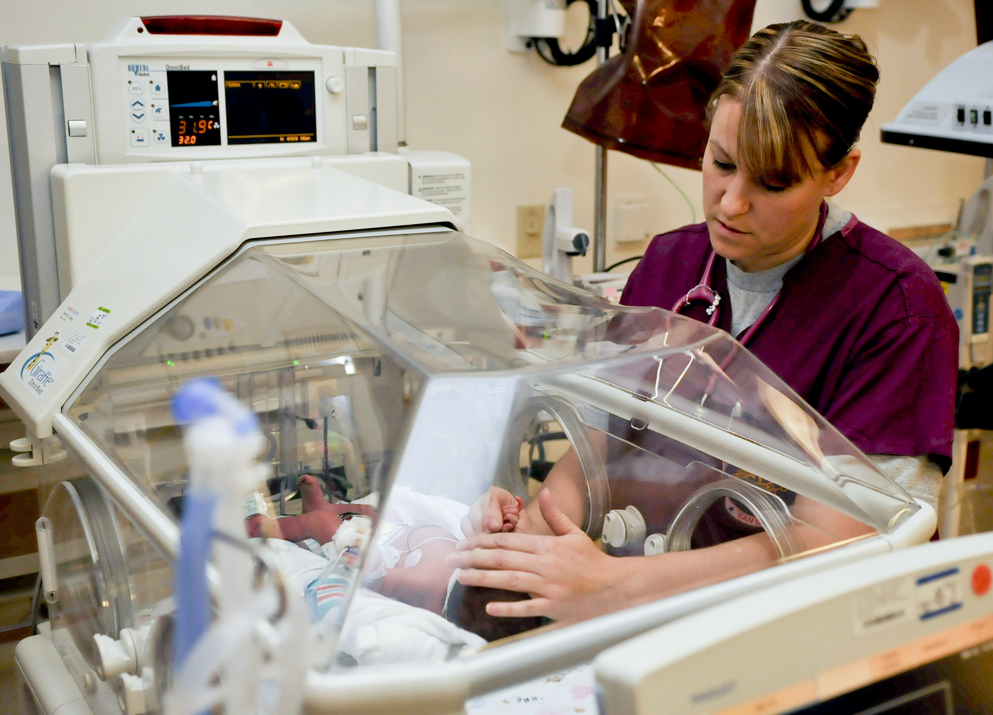
Making bread is an art, science and passion project for Fiona Strouts, PhD, a Stanford research scientist in infectious diseases.

Her baking began as a hobby several years ago, but now Strouts operates a business, L’atelier du Pain, and sells her whole-grain bread at the Portola Valley Farmers Market. I exchanged emails with her recently about her work as a professional baker and Stanford researcher.

How did you start baking bread?

“I learned to make bread about eight years ago from my Italian housemate when I lived in London during graduate school. She taught me to make 100 percent whole-wheat sourdough bread that we would bake together on the weekends. The bread was fairly dense, and provided good fuel for cycling.

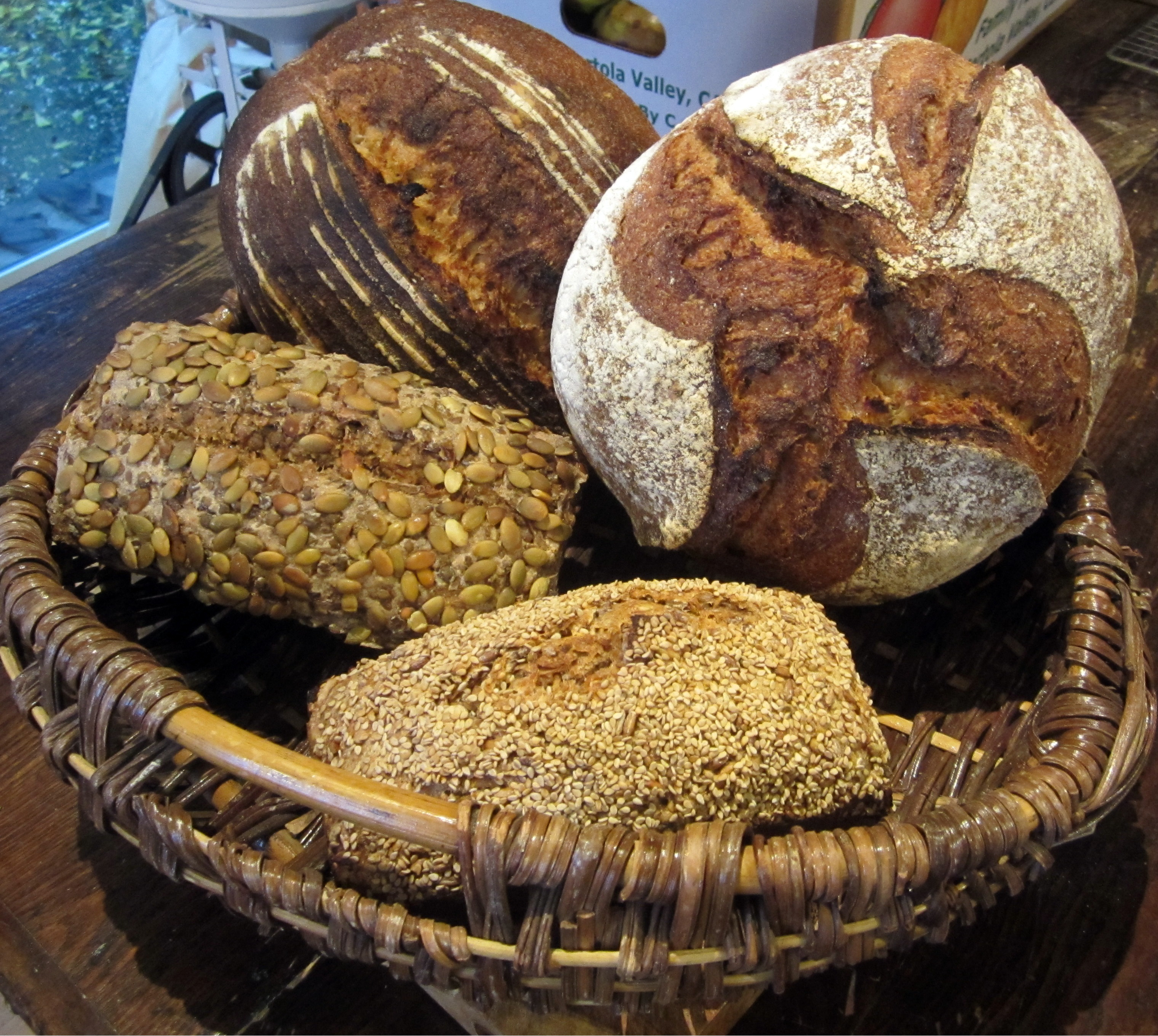
I now make whole-grain, naturally leavened breads using mostly California-grown wheat. The favorite seems to be the Sprouted Lentil & Rye bread. But my personal favorite for every-day eating is the Sonora Field Blend; it has great flavor and aroma. Sonora wheat was one of the first varieties planted in California in the early 1800s.” 

I’ve heard that you grind your own wheat. Why?

“Yes, I stone-grind my own wheat because I want to capture the flavor and nutrients, which come mostly from the germ and bran portions of the wheat berry. I buy bags of wheat berries directly from farmers, and then mill them into flour right before I mix the dough. Milling the wheat myself also ensures that the flour is 100 percent whole grain. Wheat is very nutrient-dense compared with other grains, but only when it is in the truly whole-grain form — nothing added and nothing removed from the original wheat grain.”

Why did you decide to turn your hobby into a business?

“A number of things inspired me, and they all came together a few months ago. I grew up in France, and in the village where my parents live there was a local baker and friend. The highlight of the week was going to the market on Saturday and then stopping by his house to pick up bread. There would be others from the village there and we’d share a savory pastry and a glass of wine before picking up the bread and going home for lunch. I miss that sense of community and I wanted to re-create something similar.

Then, almost a year ago I started learning more about all of the farmers in California who are passionate about sustainable agriculture and who are growing different varieties of wheat —both ancient and modern. I loved discovering the different flavors and properties of these wheats for bread making.

In addition, I’ve always been very interested in health and population health. Making whole-grain, naturally leavened breads is a way to provide a healthy option for people.”

How do you juggle baking, running a business and doing research?

“Good question! It takes organization and prioritization. I used to bike race, and the training required a lot of discipline. But starting the business was less structured and it took longer than I thought it would, as I was doing it in my spare time. I spent several weekends practicing baking large batches of bread and sharing it with some of my labmates, which I think they appreciated. The market is one day per week and it’s a manageable scale for one person. I’ve reduced my full-time equivalent [work] hours accordingly to be able to do both and my advisor has been very supportive.”

Explain your research at Stanford. Has it given you any insights into bread making?

“I work in the lab of David Relman, MD, on a project focused on improving the diagnosis and prognosis of systemic infections in humans, using sequencing of both microbial nucleic acids and host transcripts derived from blood. I am trying to understand what those blood profiles look like during states of health. And whether we’re able to detect the presence of bacteria in the blood of healthy people, to help interpret what we see in sick individuals with suspected infections.

My background has helped me understand sourdough bread making from the aspect of microbial fermentation and the effects of time and temperature. I’ve actually become quite a keen home fermenter. I have various other projects going — including yoghurt, kefir, kombucha, shoyu and miso — for which I converted the dishwasher into a fermentation chamber with a little space heater. Both baking and cooking are science, so it has also helped more generally in figuring out the properties of different types of wheat. I run a lot of bread experiments at home!”

This is a reposting of my Scope blog story, courtesy of Stanford School of Medicine.

How primary care physicians can embrace population health

Photo by Guillaume
Photo by Guillaume

The Centers for Medicare & Medicaid Services’ new payment model, called the Quality Payment Program, issues a powerful challenge to primary care physicians: Improve the quality of care and save money, by focusing on population health.

What, exactly, does that mean? In part, it means that physicians will need to consider why some groups of people are healthy and why others are not — so they make care decisions that improve the health of many people.

It sounds daunting. However, it is vital, said J. Lloyd Michener, MD, professor and chair of the Duke Department of Community and Family Medicine in a recent article in Medical Economics. 

“Small practice physicians must participate in population health; it’s the future and it’s how they’re going to get paid,” Michener said. “It’s going to be really hard not to participate in this model of care.”

There are some straightforward steps physicians can take, he said, urging physicians to:

  • Build partnerships with other care providers and health-care systems
  • Ensure electronic health records, claims data systems and other software tools capture the necessary data such as patient identification and provider-performance measures
  • Look for patterns in patient care

Population health management programs use software tools to aggregate patient data and provide a comprehensive clinical picture of each patient. Physicians then use the data to track and hopefully improve clinical outcomes while lowering costs.

For example, a primary care physician could look at all of her asthma patients that aren’t getting better, identifying how often they’ve been to the emergency room and why. This might lead to the realization that her patients are mixing up their prescription medications, so she needs to educate them further about when to use a rescue inhaler verses control inhaler. Or maybe the frequent ER visitors all live near each other, and there is an environmental issue.

Michener offered words of reassurance as well:

“Primary care physicians need to know that they have the power to quickly analyze groups of patient data and intervene when necessary to make better treatment and care decisions that lower the cost of care, improve outcomes, and raise their ability to earn income under a value-based payment system. That’s an incredibly effective way to deliver care.”

This is a reposting of my Scope blog story, courtesy of Stanford School of Medicine.

New models may help predict diabetes progression

Photo by InfoWire.dk

Diabetics exposed to consistently high blood glucose levels can develop serious secondary complications, including heart disease, stroke, blindness, kidney failure and ulcers that require the amputation of toes, feet or legs.

In order to predict which diabetic patients have a high risk for these complications, physicians may use mathematical models. For example, the UKPDS Risk Engine calculates a diabetic patient’s risk of coronary heart disease and stroke — based on their age, sex, ethnicity, smoking status, time since diabetes diagnosis and other variables.

But this strategy doesn’t provide the accuracy needed by doctors. In response, a research team at Duke University has developed machine-learning computer algorithms to search for patterns and correlations in EHR data from approximately 17,000 diabetic patients in the Duke health system.

The group, led by Ricardo Henao, an assistant research professor in electrical and computer engineering, has demonstrated more accurate predictions than the UKPDS Risk Engine. A recent news story explains:

“This new model can project whether a patient will require amputation within a year with almost 90 percent accuracy, and can correctly predict the risks of coronary artery disease, heart failure and kidney disease in four out of five cases. The model looks at what was typed into a patient’s chart — diagnosis codes, medications, laboratory tests — and picks up on which pieces of information in the EHR are correlated with the development of a comorbidity in the following year.”

The Duke researchers plan to improve the model by training their machine-learning algorithms on a larger data set of diabetic patients from additional hospitals.

However, relying on EHR data has drawbacks. For instance, a patient’s EHR may be incomplete, particularly if the patient doesn’t consistently see the same doctors. Another major challenge is gaining access to the medical records for research. The Duke team had to contact all 17,000 patients to get their informed consent and may encounter similar challenges for a larger scale project.

This is a reposting of my Scope blog story, courtesy of Stanford School of Medicine.

How does burnout affect NICU caregivers and their patients?

a_nurse_examines_a_newborn_baby
Photo by U.S. Navy

We’ve all felt burned out at work due to prolonged stress — physically and emotionally exhausted, unmotivated, frustrated and maybe even cynical. But in the health-care field, burnout can harm patients as well as workers.

That concern prompted Stanford researchers to investigate the prevalence of caregiver burnout in neonatal intensive care units (NICUs) to determine whether it is correlated with healthcare-associated infections. The study is reported in the Journal of Perinatology.

The research team analyzed survey data from the California Perinatal Quality Care Collaborative, including responses from over 2000 providers — physicians, nurses and other workers — in 44 California NICUs who cared for over 4000 very low birth rate infants. One quarter of the respondents reported symptoms consistent with burnout on average, but burnout prevalence varied from 7.5 to 54.4 percent within each NICU.

I spoke recently with the study’s lead author, Daniel Tawfik, MD, a critical care fellow at Lucile Packard Children’s Hospital.

What inspired you to investigate caregiver burnout in NICUs?

“Throughout my medical training, I received very little education on the concepts of mindfulness and medical provider well being. But the challenging experiences and stressful situations encountered every day in the hospital must have some impact on the providers’ mental health and the care they deliver to their patients. This study was a way to evaluate this relationship and hopefully spur greater awareness of burnout and encourage research to address its role in patient care.”

What did you find? Were there any surprises?

“We expected to find increased healthcare-associated infections in NICUs with high levels of burnout, since preventing infections in these vulnerable patients takes a great deal of vigilance.

We were a little surprised that there wasn’t a strong relationship apparent when we analyzed the data. This may be due to the relatively small number of NICUs and the fact that infections were fortunately not very common in this group of infants.

Although our most recent analyses show increased burnout among large NICUs, our research group previously reported improved outcomes among large NICUs. It’s possible that the largest NICUs have increased burnout, but they also have systems in place to improve the quality of care — counteracting the effect that burnout may have on quality indicators.

We were also a little surprised to find that physician burnout [approximately 17 percent] was less prevalent than non-physician burnout [approximately 28 percent]. But this discrepancy is also in line with our previous studies in which nurses reported lower ratings of safety culture and a desire for more respect and input in decision-making. It’s possible that these differences contributed to increased burnout among nurses, who made up the majority of our survey respondents in our new study.”

How can your results be used to improve NICU care?

“These results highlight the prevalence of burnout among NICU providers, particularly among nurses. Whether or not this burnout affects quality of care in measurable ways, it suggests that we need interventions to prevent and reduce burnout in NICUs and likely in other care settings.

The NICU at Lucile Packard Children’s Hospital is one of several NICUs around the country participating in the WISER randomized-controlled trial, which is evaluating the effectiveness of burnout interventions such as daily recall of positive events using the Three Good Things tool. The study is being led by Jochen Profit, MD, and Bryan Sexton, PhD, who have been my mentors and co-authors. It’s our hope that if this intervention proves beneficial that it could be expanded to other hospital units and clinics.”

This is a reposting of my Scope blog story, courtesy of Stanford School of Medicine.

Saliva tests may help identify marijuana-impaired drivers

Photo by ashton
Photo by ashton

As of the recent election, seven states and the District of Columbia have now legalized marijuana for recreational use and 19 other states have legalized medical marijuana. And this legalization has raised concerns about driving under the influence of marijuana.

A number of research groups are now focusing on ways to identify drivers impaired by marijuana. As recently reported by KQED, the Center of Medicinal Cannabis Research at the University of California, San Diego, are working to “gather data about dosages, time and what it takes to impair driving ability — and then create a viable roadside sobriety test for cannabis.” And a group of Stanford engineers have created a test called a ‘potalyzer.’

The Stanford effort was led by Shan Wang, PhD, a Stanford professor of materials science and engineering and of electrical engineering. He and his colleagues developed a mobile device that detects the amount of tetrahydrocannabinol (THC) molecules in saliva. (THC is the main psychoactive agent in marijuana.)

The test would allow police officers to collect a saliva sample from the driver’s mouth with a cotton swab, analyze it with the new device, and then read the results on a smartphone or laptop in as little as three minutes.

The technology combines magnetic nanotechnology with a competitive immunoassay. During the test, saliva is mixed with antibodies that bind to both THC molecules and magnetic nanoparticles. The mixture is placed on a disposable test chip, inserted into the handheld device and the THC-antibody-nanoparticles are detected by magnetic biosensors. The biosensor signal is then displayed on a Bluetooth-enabled device.

Wang’s group focused on developing a THC saliva test because it is less invasive and may correlate better with impairment than THC urine or blood tests. Also key is the need for a very sensitive test. A Stanford news release explains:

“Wang’s device can detect concentrations of THC in the range of 0 to 50 nanograms per milliliter of saliva. While there’s no consensus on how much THC in a driver’s system is too much, previous studies have suggested a cutoff between 2 and 25 ng/ml, well within the capability of Wang’s device.”

There is still a lot to do before police can deploy this ‘potalyzer’ device, including making it more user-friendly, getting it approved by regulators and investigating whether there is a better biomarker to detect marijuana impairment than THC. In addition, the test may not work well for THC edibles, the researchers wrote in a recent paper published in Analytical Chemistry.

On the upside, the Stanford technology could also be used to test for morphine, heroin, cocaine or other drugs — and for multiple drugs at the same time.

More research is needed, but there is now a new funding source in California: Proposition 64 allots millions of dollars per year to research marijuana and develop ways to identify impaired drivers.

This is an expanded version of my Scope blog story, courtesy of Stanford School of Medicine.

Many adults should now take statins, task force recommends

Illustration by Rf Vectorscom

The U.S. Preventative Services Task Force now recommends that many adults take a low to moderate dose of statin to reduce their risk of a heart attack or stroke, even if they don’t have a history of cardiovascular disease.

Statins are drugs that reduce the production of cholesterol by the liver — lowering bad cholesterol and triglycerides and raising good cholesterol. The task force comprehensively reviewed the literature on clinical trials and observational studies involving statin use. It concluded that the benefits of using statins outweighed the harms in some patients with increased risk of cardiovascular disease.

Douglas Owen, MD, a Stanford professor of medicine and director of the Center for Health Policy, was a member of the task force when the guidelines were developed. He summarizes the new recommendations in a recent news story:

“The task force recommends that clinicians offer statins to adults who are 40 to 75 years old and have at least one existing cardiovascular disease risk, such as diabetes, hypertension, high cholesterol or smoking. They also must have a calculated risk of 10 percent or more that they will experience a heart attack or stroke in the next decade. The task force recommends that clinicians use the American College of Cardiology/American Heart Association risk calculator to estimate cardiovascular risk because it provides gender- and race-specific estimates of heart disease and stroke.”

The task force hope these new recommendations will help clinicians better identify cardiovascular risk, so their patients can take steps to reduce their risk, such as eating a healthy diet, exercising and potentially taking a statin.

This is a reposting of my Scope blog story, courtesy of Stanford School of Medicine.

Brain scans detect lies better than polygraph tests, new study shows

8742707669_786cfa376b_k
Photo by Tristan Schmurr

Forget fact checkers or polygraph tests. A functional magnetic resonance imaging (fMRI) brain scan might be the best way to tell if someone is lying.

According to a study from the University of Pennsylvania, our brains are more likely to give us away when we’re lying than sweaty palms, rapid breathing or spikes in blood pressure, the factors tracked by polygraph tests.

The researchers directly compared the ability of two techniques — fMRI and polygraph tests — to detect concealed information. They had 28 participants secretly write down a number between 3 and 8 on a slip of paper. Each participant then had both lie-detection tests, in random order, a few hours apart. During both sessions, they always answered “no” when asked if they had picked a certain number, which meant that one out of the six answers was a lie.

Three fMRI experts and three professional polygraph examiners then independently analyzed the results. The fMRI experts were 24 percent more likely to detect the lie than the polygraph experts, as recently reported in the Journal of Clinical Psychiatry.

Although the study wasn’t designed to evaluate the combined use of both techniques, the polygraph and fMRI results agreed correctly on the concealed number for 17 participants. So they plan to investigate in the future whether these techniques are complementary.

The study includes only a small number of participants, but the research team is encouraged by the results. “While the jury remains out on whether fMRI will ever become a forensic tool, these data certainly justify further investigation of its potential,” said Daniel Langleben, MD, first author and a professor in psychiatry, in a recent news release.

This is a reposting of my Scope blog story, courtesy of Stanford School of Medicine.

Talk about death — before a health crises, says Stanford’s Philip Pizzo

walk-842535_1280
Photo by Oldiefan

Most of us have no desire to think, or talk, about death. However, if we never talk about it, we leave our health providers and families guessing about how and where we want to die.

That’s why it’s important to communicate end-of-life preferences early, Philip Pizzo, MD, founder and director of the Stanford Distinguished Careers Institute, argues in a recent perspective in the Proceedings of the National Academy of Sciences.

It’s a topic he’s quite familiar with: Pizzo co-authored the Institute of Medicine’s report “Dying in America,” which addressed how to improve the quality of care for patients with advanced illnesses, without exacerbating the high costs of health care. In the perspective, he summarizes the IOM report’s key recommendations and provides an update.

Pizzo discussed the piece in an email:

“Unless we are facing an illness or event that makes the prospect of death imminent, most of us do not even think about the inevitability of our mortality. These conversations get slotted to times when death is more imminent and when our crisis-oriented decisions may not reflect our true preferences. That is why the IOM report recommended that conversations about death take place with our health care providers and families throughout life. Our thoughts and preferences about dying will vary at different stages of life and wellbeing.”

In the perspective, Pizzo describes the progress that has been made since the report was published. For instance, the Centers for Medicare & Medicaid Services in January 2016 began paying doctors to have end-of-life conversations with patients — a move Pizzo lauds as a major step.

Another important achievement, according to Pizzo, is the national stakeholder conferences that are now bringing constituencies together to implement the report’s recommendations.

He added:

“We witnessed before the IOM Committee began its work how rapidly public opinion can be swayed by political rhetoric. Thankfully since then, the public’s willingness to engage in conversations about death and dying have become better realized and books, like Atul Gawande’s Being Mortal or Paul Kalaniti’s When Breath Becomes Air, have helped to foster more enlightened conversations about dying.”

This is a reposting of my Scope blog story, courtesy of Stanford School of Medicine.

Alzheimer’s researchers call on citizen scientists to play an online game

Image by geralt
Image by geralt

Many people, like me, have helplessly watched a loved one suffer and die from dementia. Now there is something you can do to help accelerate Alzheimer’s research — play a game.

The game, called Stall Catchers, is part of the EyesOnALZ project that uses citizen scientists to analyze Alzheimer’s research data. The game was developed by the Human Computation Institute, in collaboration with scientists from Cornell University, MIT and the University of California, Berkeley. The research team is trying to understand the association between reduced blood flow in the brain and Alzheimer’s disease.

The game features movies of real blood vessels in live mouse brains. Players must search for clogged vessels where blood flow is blocked, or stalled. Each movie is seen by many citizen scientists and then checked by a research scientist in order to quickly and accurately identify the stalls.

Past research has shown that Alzheimer’s is associated with the accumulation of beta amyloid proteins that clump together into sticky, neurotoxic aggregates called amyloid plaques. These proteins are normally cleared by the blood stream, but the formation of amyloid plaques slows down this clearance process.

Recent animal studies, performed by the Schaffer-Nishimura Lab at Cornell, suggest that improving blood flow in the brain may help reduce the devastating effects of amyloid accumulation. The researchers discovered that up to two percent of capillaries in the brains of Alzhiemer’s-affected mice were clogged — 10 times more than usual — and this caused up to a 30 percent decrease in overall blood flow in the brain.

“Advanced optical techniques have allowed us to peer into the brain of mice affected by Alzheimer’s disease,” said Chris Schaffer, PhD, the principal investigator in the Schaffer-Nishimura Lab, in a recent news release. “For the first time, we were able to identify the mechanism that is responsible for the significant blood flow reduction in Alzheimer’s, and were even able to reverse some of the cognitive symptoms typical to the disease.”

Now the main challenge for the Cornell researchers is the time-consuming process of manually analyzing all the brain movies to identify the stalled vessels. They need to study up to a thousand vessels for each animal. That’s why they collaborated with the experienced citizen teams at UC Berkeley and MIT to create the Stall Catchers game to get help from the public.

“Today, we have a handful of lab experts putting their eyes on the research data,” said Pietro Michelucci, PhD, the EyesOnALZ principal investigator, in a news story. “If we can enlist thousands of people to do that same analysis by playing an online game, then we have created a huge force multiplier in our fight against this dreadful disease.”

This is a reposting of my Scope blog story, courtesy of Stanford School of Medicine.


Researchers discover “brain signature” for fibromyalgia using brain scans

portrait-1006703_1280Millions of patients suffering from fibromyalgia often experience widespread musculoskeletal pain, sleep disturbances, fatigue, headaches and mood disorders. Many also struggle to even get diagnosed, since there are currently no laboratory tests for fibromyalgia and the main symptoms overlap with many other conditions. However, new research may help.

Scientists from the University of Colorado, Boulder may have found a pattern of brain activity that identifies the disease. They used functional MRI (fMRI) scans to study the brain activity of 37 fibromyalgia patients and 35 matched healthy controls, while the participants were exposed to a series of painful and non-painful sensations.

As reported recently in the journal PAIN, the research team identified three specific neurological patterns correlated with fibromyalgia patients’ hypersensitivity to pain.

Using the combination of all three patterns, they were able to correctly classify the fibromyalgia patients and the controls with 92 percent sensitivity and 94 percent specificity — meaning that their test accurately identified 92 percent of those with and 94 percent of those without the disease.

Tor Wager, PhD, senior author and director of the school’s Cognitive and Affective Control Laboratory, explained the significance of the work in a recent news release:

“Though many pain specialists have established clinical procedures for diagnosing fibromyalgia, the clinical label does not explain what is happening neurologically and it does not reflect the full individuality of patients’ suffering. The potential for brain measures like the ones we developed here is that they can tell us something about the particular brain abnormalities that drive an individual’s suffering. That can help us both recognize fibromyalgia for what it is – a disorder of the central nervous system – and treat it more effectively.”

More research is needed, but this study sheds a bit of light on this “invisible” disease.

This is a reposting of my Scope blog story, courtesy of Stanford School of Medicine.