Berkeley Physics was delighted to host the 2023 Rising Stars in Physics Workshop this spring, sponsored by the Heising-Simons Foundation. This event brought together 24 outstanding women physicists and astronomers for two days of scientific talks and informal discussions aimed at helping them navigate the early stage of their academic careers.
The workshop was led by Pablo Jarillo-Herrero, associate professor of physics at MIT and founder of the workshop series, and Alessandra Lanzara, professor and Charles Kittel Chair in Physics at UC Berkeley. Lanzara’s personal experiences motivated her to help organize it. “My first years at Berkeley were difficult with too few role models in my department. I often felt lonely and like I didn’t belong,” says Lanzara. “These types of workshops help create a network for young women to share experiences, challenges, and ideas with their peers and senior colleagues to help them succeed.”
The highlight of the workshop for many of the participants was getting to know each other—through workshop sessions and casual interactions over meals or in an airport shuttle. “Meeting my physics peers from different fields was amazing,” says Veronika Sunko, Miller Postdoctoral Fellow. “The atmosphere was different with 80% of the people in the room being women. It was fun.” Sunko is a condensed matter experimentalist studying quantum materials with Berkeley Physics Professors Joe Orenstein and James Analytis. “We use lasers to learn about the magnetism of materials that exhibit an interesting interplay of magnetic and electronic degrees of freedom, yielding new phenomena that we’re working to understand,” she says.
Weishuang (Linda) Xu, postdoctoral researcher at the Berkeley Center for Theoretical Physics, enjoyed the keynote speeches by senior investigators. “Very accomplished people talked in-depth about their career trajectory and the massive-scale projects they’re overseeing,” says Xu. “I also appreciated the participants’ talks. I rarely hear about research outside my narrow niche of theoretical particle physics.” Xu works on various projects with different collaborators, using astrophysics and cosmology tools to study the particle nature of dark matter. “For example, I collaborate with Berkeley Physics Assistant Professor Ben Safdi. We’re analyzing data of gamma rays that come from the middle of our galaxy to search for Higgsino dark matter,” explains Xu.
As a new postdoc at the Harvard Society of Fellows, Carolyn Zhang soaked in career tips from workshop panelists on how to apply for faculty positions, build a research group, and balance commitments. “It was helpful to hear about different people’s unique journeys to where they are today. And it was cool to see so many women physics professors in one room,” she says. As a condensed matter theorist, Zhang studies the quantum phases of matter and the transitions between them. But her interests are broad and her fellowship is not tied to a single department.
Zhang also appreciates the volunteers who devoted time to organize the workshop. “They seemed genuinely passionate about supporting women entering the physics field, which was very encouraging.”
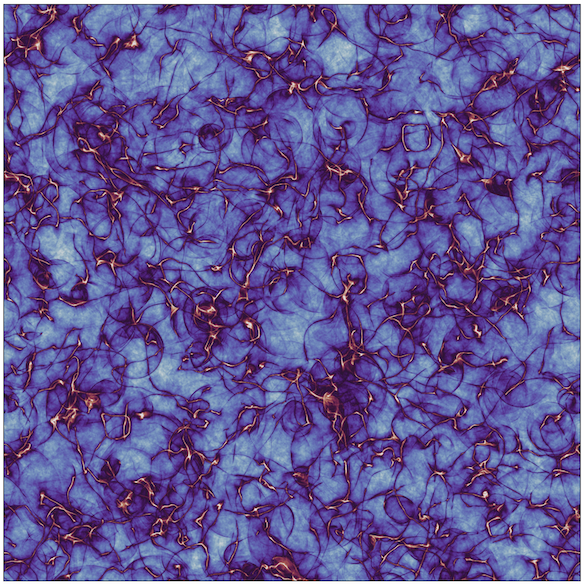
A zoom-in of axion strings in a simulation of axion dark matter in the early universe. The strings throw off axions that go on to become that dark matter. These simulations are used to predict the properties of the axion, which informs laboratory detection efforts. (Ben Sadfi, UC Berkeley)
SCIENTISTS HAVE BEEN STUDYING THE COSMOS for centuries, but we still don’t know what makes up 85% of all matter in the universe. Unlike ordinary matter that we can see and feel, dark matter hasn’t been observed directly by even our most advanced scientific instruments. These invisible particles may be zipping through us all the time without interacting.
But scientists believe our world wouldn’t exist without dark matter. Its gravitational pull holds galaxies together, gathers them into clusters, bends light around them, and affects how they rotate. Dark matter also played a crucial role as galaxies initially formed.
“Lots of observational data show us that dark matter is a real particle, but we don’t know what kind. Its possible mass has a huge range, and there might be multiple types of dark matter particles,” says Berkeley Physics Professor Dan McKinsey, the Georgia Lee Chair in Physics. “We’re working hard to detect dark matter in the lab to open a window into new physics. It’s the only particle we know to exist outside the standard model.”
Berkeley Physics is one of the top places in the world to study dark matter. Experimental and theoretical physicists at Berkeley are leading far-reaching searches—hunting for dark matter candidates ranging from 1 TeV weakly interactive massive particles (WIMPs) to 1 MeV light dark matter particles down to 10 μeV axions.
Berkeley faculty are conducting, building, and designing next-generation dark matter experiments, including the LZ, SuperCDMS, TESSERACT, and ALPHA plasma haloscope. These innovative experiments are guided by models developed by Berkeley Physics theorists, including professors Hitoshi Murayama, Lawrence Hall, and Ben Safdi. We highlight only a few of these comprehensive efforts here.
HUNTING FOR WIMPS WITH LZ
One promising candidate is WIMPs, weakly interacting but heavy dark matter particles with a predicted mass of about 10 GeV to 100 TeV. A GeV is roughly the mass of a proton.
Hunting for WIMPs over 9 GeV is the aim of LZ, the larger and more sensitive successor of the LUX experiment. After 60 days of running, LZ recently became the most sensitive dark matter detector in the world. Berkeley Physics Professor and Berkeley Lab Director Mike Witherell, Emeritus Professor Bob Jacobsen, and McKinsey contributed to this success.
Because dark matter particles rarely interact with ordinary matter, their signal is easily drowned out by background noise. To shield from cosmic rays, LZ is located nearly a mile underground at the Sanford Underground Research Facility (SURF) in South Dakota. To reduce radioactive contamination, it uses ultra-clean detector materials. And to lower environmental backgrounds, it is built in several layers like an onion.
At the center of LZ is a time projection chamber (TPC)—a tank filled with seven tons of highly-purified liquid xenon. If a dark matter particle strikes a xenon nucleus, a flash of light and an electric charge are produced as the nucleus recoils. A strong electric field drifts the charge to the top surface of the TPC, where the electrons create a much larger flash of light that is measured by photomultiplier tubes on top and bottom.
The pattern and timing of the two flashes pinpoint the position and energy of the event. And the ratio of the two scintillation signals determines if the event was caused by a nuclear or electron recoil.
Outside the TPC are two veto detectors—a “skin” hold- ing three tons of liquid xenon and then an “outer detector” of gadolinium-loaded liquid scintillator—which are used to reject signals from gamma rays and neutrons, respectively. The whole thing lives inside a massive tank of water.
LZ is 25 times larger than the previous generation LUX experiment, which helps suppress backgrounds. But this increase also created a major challenge for McKinsey: designing and building a much higher high-voltage system to get the correct drift electric field, without the xenon lighting up like a neon lamp.
McKinsey also led the data analysis effort to reduce “accidental backgrounds” with support on backgrounds from Berkeley Physics postdoc Ibles Olcina and graduate students Jose Soria, Yue Wang, Ryan Gibbons, Ryan Smith, and James “Reed” Watson. “Occasionally, isolated first and second scintillation pulses randomly pair up to look like a dark matter event,” explains McKinsey. “My group combed through data, produced a statistical model, and developed cuts to reduce these accidentals without cutting into our dark matter acceptance.”
So far, LZ has found no evidence of WIMPs, but it set the most stringent limits on WIMP cross-sections and masses to date. And the second 1000-day run is underway.
“LZ is performing to specification, which is a big deal since we’ve been working on it for a decade,” says McKinsey. “We’re now poised to push through more dark matter parameter space over the next few years.”
McKinsey is also helping to design the next-generation of LZ, a scaled-up 80-ton xenon experiment called XLZD.
SEARCHING FOR LOW-MASS WIMPS WITH SUPERCDMS
SuperCDMS, the next-generation of the CDMS experiment, is located deep underground at SNOLAB near Sudbury, Canada. It plans to detect dark matter particles with a mass between 10 GeV and 0.5 GeV. Berkeley Physics Professor Emeritus Bernard Sadoulet led the NSF-funded part of its construction.
Searching for dark matter with lower mass requires more sensitive detectors. SuperCDMS uses germanium or silicon crystals attached to sensors on both faces. When a dark matter particle interacts with either semi-conductor, its nucleus recoils and creates minute crystal vibrations (phonons) and ionization (charge). An electric field causes the charge to drift and shed lots of phonons.
“Measuring both phonons and ionization gives us discrimination capability against backgrounds. And drifting the charges in the high-voltage detector increases our energy sensitivity by a factor of 100, allowing us to search for lower masses,” says Sadoulet.
However, measuring these phonon signals is challenging. Berkeley Physics Assistant Professor Matt Pyle played a major role in developing this unique sensor technology with the help of Berkeley’s associate research physicist Bruno Serfass and former postdoctoral fellow William Page.
At its core are transition-edge sensors (TES)—materials stabilized in the middle of their superconducting transition—attached to aluminum fin antennas. The fin absorbs the energy of the vibrations, concentrates it, and pushes it into the TES. The resulting increase in TES temperature changes its resistance, which is measured by cryogenic electronics.
“These phonon sensors need to be small to reduce their heat capacity. But if they’re attached directly to a giant crystal, an athermal phonon bounces around for a long time before it interacts with the TES. By using fins, we increase the interaction probability and area coverage,” says Pyle, the Michael M. Garland Chair in Physics.
He adds, “Only about 30% of the energy is transferred to the TES using the fins, but that’s more than made up for by collecting the phonons quickly before they thermalize.”
The collaboration is currently installing the experiment at SNOLAB. Meanwhile, they have been commissioning the SuperCDMS detectors, software, and operations at CUTE, a nearby cryogenic underground test facility at SNOLAB.
Sadoulet notes that Berkeley Physics collaborations encourage technology and data analysis transfer between various groups. The core athermal phonon sensor technology and discrimination methods are being used in multiple experiments, including SuperCDMS and TESSERACT.
“We’re giving a single solution that will hopefully be employed many times by many different experiments, all searching for slightly different things,” says Pyle.
SEEKING LIGHT DARK MATTER WITH TESSERACT
TESSERACT intends to take the dark matter search a step further. This umbrella of two experiments is being designed to detect light dark matter particles from both nuclear and electron recoils, in the mass range of the proton to the electron—1 GeV down to 1 keV.
The entire project will use identical Berkeley Physics next-generation sensors, readout technology, and operations—and no electric field for signal amplification.
“What makes TESSERACT unique is that every detector is designed to have multiple signal channels that have to be in coincidence for dark matter events. That’s the secret idea sauce of TESSERACT,” says Pyle. “We’re also eliminating a whole class of backgrounds by going to zero field, which means we need very highly sensitive detectors.”
McKinsey’s group is helping develop TESSERACT’s HeRALD experiment with assistance from Assistant Project Scientist Junsong Lin and graduate students Roger Romani, Will Matava, and Wang. HeRALD uses purified liquid helium as the target for nuclear recoil dark matter. Its silicon athermal phonon detectors are submerged in the vat of liquid helium and suspended in a vacuum above it.
“Using helium provides excellent background discrimination. If a dark matter event occurs in the helium, it lights up multiple pixels in coincidence, whereas a background event or microfracture in the silicon only lights up one pixel.” Helium is also cheap, easy to purify, easy to scale up, and naturally immune to some backgrounds. HeRALD will initially be sensitive to dark matter particles from 1 GeV to 100 MeV, but the scientists hope to reach the keV scale in the future.
Pyle’s group is helping develop TESSERACT’s SPICE experiment. It uses polar crystals—either gallium arsenide (GaAs) or sapphire (Al2O3)—as the target for both nuclear and electron recoil dark matter. A polar crystal has two types of ions with opposite charges. Some dark matter candidates may transform themselves, with low probability, into photons, which then nudge the different ions in opposite directions. This produces phonons that can be detected by the TES.
For the GaAs part of SPICE, the photons and phonons are collected in separate detectors, enabling photon-phonon coincidence to tag the unique dark matter signature, says Pyle. This scheme is designed to detect dark matter between 1 GeV to 1 MeV, caused primarily by electron scattering.
To detect dark matter with even lower mass, the second part of SPICE measures only the athermal phonon signal using sapphire detectors with even better energy sensitivity.
Pyle’s group is unraveling how to read out and calibrate these sapphire crystalline targets using his latest TES. Currently, the detectors are sensitive to dark matter in the GeV to MeV range, but the team hopes to get down to a keV.
McKinsey and Pyle are both enjoying their tabletop experiments. “If the detector is sensitive enough, then you can in principle detect light dark matter in your lab at the surface. It could happen,” McKinsey says. But TESSERACT will be installed underground in the next few years—at SURF or the Modane Underground Laboratory.
PROBING FOR AXIONS
Berkeley Physics Assistant Professor Ben Safdi studies other dark matter candidates, but he finds the ultra-light axions to be the most compelling because they explain more than dark matter. Axions were first theorized to explain the mystery of how neutrons behave in electric fields. And they have deep connections to the consistency of gravity at a quantum level.
“Axions are theoretically very well motivated, and they’re almost completely unexplored experimentally,” says Safdi, who holds the Henry Shenker Professor in Physics. “In the next decade or so, we’ll be able to say definitively whether or not these particles exist in nature.”
Although Safdi is a theoretical physicist, he looks for indirect signatures of dark matter in experimental data with his team, including graduate students Yujin Park and Joshua Benabou. “My work starts theoretically, with pencil and paper and then simulations. For a particular astrophysical system or precision laboratory experiment, we ask how axions would affect it and what data we need to test these predictions. Then, we get and analyze those data sets, determining if we find evidence for axions or not,” explains Safdi.
Additionally, Safdi spends much of his time simulating how axions were produced shortly after the Big Bang to determine what mass gives the correct abundance of dark matter. For this work, he uses advanced supercomputers at the National Energy Research Scientific Computing Center (NERSC) at Berkeley Lab, which just commissioned its faster and bigger Perlmutter supercomputer. “We have lots of jobs up the hill churning away. It’s a game changer for our mass prediction computations,” says Safdi.
One feature he needs to simulate in the early universe is axion strings, which are very violent but narrow regions of space—like tiny tornadoes—that whip around and emit lots of axions.
“During the simulations, a small part of the expanding universe is represented by a 3D grid over which the equations are solved,” explains Safdi. “But the axion strings are moving, so we have to dynamically update the grid. Despite running on supercomputers, computer memory is our limiting factor.”
Luckily, Safdi teamed up with the AMReX collaboration at Berkeley Lab, adapting their code framework designed to solve multi-scale problems. The key was using an adaptive mesh grid with a fine spatial resolution around the axion strings and sparse resolution elsewhere.
Using the biggest simulation of cosmology to date, they more precisely predicted the axion mass to be between 180 to 40 μeV, higher than expected. This claim implies axions from the early universe can’t be detected by the current experiments, which use a microwave resonance chamber to enhance the photon frequency coming from axions. The required chamber would be too small to get a measurable signal.
However, Safdi’s prediction excited Berkeley Nuclear Engineering Professor Karl van Bibber. He is building the ALPHA plasmonic haloscope, which creates resonant enhancement using parallel wires in a strong magnetic field. And van Bibber is waiting to tune his experiment using the more precise predictions Safdi is now calculating with the Perlmutter.
Overall, Berkeley Physics’ search for dark matter is casting an impressively wide net. “Berkeley might be the best place in the world for dark matter research. I can’t think of any place that’s stronger overall and better,” says McKinsey.
This is a reposting of my Berkeley Physics magazine feature, courtesy of UC Berkeley’s Physics Department.
Ashley James is fascinated by her toxicology research because it combines biology, chemistry, physics and morbid topics like poisonings that affect the environment and world health. She also loves the unexpected twists. “We’ve been surprised so many times by what we’ve found. It’s been a fun, wild ride,” she said.
As a PhD student and now postdoctoral research fellow from the University of Saskatchewan, James’ research on mercury poisoning in animals and humans uses X-rays produced by the Stanford Synchrotron Radiation Lightsource (SSRL) at the Department of Energy’s SLAC National Accelerator Laboratory.
For her work, James will receive the 2023 Melvin P. Klein Scientific Development Award during the 2023 SSRL/LCLS Annual Users’ Meeting held at SLAC September 24-29.
“The SSRL-based research of Dr. James addresses a global health question with breakthrough discoveries while demonstrating state-of-the-art methodology for her discipline, thus bearing all the hallmarks of ‘outstanding research accomplishments by a new investigator’ that the award is intended to recognize,” wrote her PhD supervisors Graham George and Ingrid Pickering, Canada Research Chairs and professors at the University of Saskatchewan, in a nomination letter for the award.
James said she felt surprised, excited and honored by SSRL when she found out about winning the award. “It’s awesome to be included in the list of Klein awardees, alongside the diverse projects and incredible scientists who’ve won in the past,” she said.
Re-thinking the Minamata mass poisoning
As a PhD student, James studied the mass poisoning of thousands of people who ingested mercury by eating tainted fish and shellfish from the Minamata Bay in Japan during the late 1950s and 1960s. This famous, deadly tragedy was caused by a chemical plant dumping mercury-contaminated industrial waste into the bay – as demonstrated by a physician working for the factory who fed cats food laced with the industrial waste to confirm that it was responsible for the neurological disease.
Because the chemical plant used inorganic mercury in its processes, scientists initially believed that the contaminated waste contained inorganic mercury. The idea was that this inorganic mercury was transformed in the environment into a common and more toxic form of organic mercury called methylmercury.
James and her collaborators investigated the Minamata poisoning by studying preserved samples from a cat in the historic study. First, they showed that the mercury in the cat’s brain tissue was mostly organic by performing studies at SSRL’s X-ray spectroscopy (XAS) beamline 7-3 and high-energy-resolution fluorescence detection (HERFD-XAS) beamline 6-2, with help from SLAC scientists Matthew Latimer, Thomas Kroll and Dimosthenis Sokaras.
“These synchrotron techniques allowed us to look at historical samples with X-ray eyes to determine what mercury compounds existed in the cat’s brain tissue, which then told us a lot about its toxicology,” explained James. “HERFD-XAS has been particularly useful because its higher resolution enhances the shape of the mercury spectra, so we can fit the complex mixture of compounds with more confidence.”
The team then used computer-based calculations to model the chemical plant’s processes. Instead of methylmercury, their computational chemistry studies predicted that the factory released a different organic mercury compound called alpha-mercuri-acetaldehyde, whose toxicology has not been studied. Their findings challenged the long-standing view of what form of mercury poisoned the human population in Minamata.
The ensuing controversy attracted media attention and some scientific criticism, which was a bit overwhelming and stressful for James as an early-career PhD student. However, she handled it like a veteran, according to her nomination letters. Her supporters described the criticism published in letters to the journal as personal and unscientific. And they praised James’ response as excellent and surgically precise.
“Science is meant to create debate and it certainly did that,” said James. “But our main point was that it is important to study the toxicology of organic mercury compounds other than methylmercury, because they may have important environmental and health impacts.”
Comparing acute and chronic mercury poisoning in humans
Her second PhD research project investigated the more prevalent issue of chronic mercury exposure due to a lifetime of eating marine fish with low levels of methylmercury, which can lead to as much as ten times higher concentrations of mercury in the brain. Specifically, she used the same X-ray spectroscopy techniques and SSRL facilities as the Minamata project to study brain samples from residents of the Seychelles islands.
She compared these results to similar beamline studies on two historic acute organic mercury poisoning cases, which involved a short-term exposure of large concentrations of organic mercury. All brain tissue samples for her PhD work were acquired through her collaborators at the University of Rochester.
“We wanted to see if there was a difference in the chemical form of mercury found in chronic versus acute human exposure cases,” said James. “Honestly, we found the complete opposite of what we expected.”
The human body uses a chemical process called demethylation as a defense mechanism to slowly turn organic mercury into less toxic inorganic mercury. The researchers therefore thought people showing no evident symptoms of mercury poisoning would have mostly inorganic mercury that had been demethylated, James explained. Instead, they found individuals with chronic exposure had low concentrations of mercury in their brains, but it was entirely organic.
Similarly, the scientists predicted individuals after acute poisoning would have less time to demethylate the mercury, meaning they would have high levels of organic and low levels of inorganic mercury. Instead, they found complex mixtures with low concentrations of organic and high concentrations of inorganic mercury.
“The takeaway of this crazy twist is that it could be misleading to use acute exposure studies to understand the vast majority of human exposures that are chronic in nature,” said James. “Over a billion people worldwide depend on fish as their primary or sole source of protein. So, better understanding the ramifications of ingesting fish that may contain low levels of mercury is an important global food security question.”
As a postdoc, James is extending her mercury toxicology research. She is now studying the role of metals in multiple sclerosis using a diverse range of SSRL X-ray spectroscopy and X-ray imaging beamlines.
“Dr. James’ remarkably impactful research is further distinguished by her use of an incredible range of different techniques — from advanced X-ray spectroscopy methods at SSRL to quantum chemistry, alongside more conventional toxicology methodology,” said George and Pickering. “This work represents one of the very first demonstrations of these techniques to her field.”
The Klein award is named in honor of the late Melvin P. Klein, a world-renowned biophysicist at Lawrence Berkeley National Laboratory and the University of California, Berkeley and a longtime user at SSRL.
Almost every day, news outlets report on highly infectious COVID-19 variants threatening to sneak past the front-line antibody defenses developed by our bodies after vaccination or previous infection. That’s because the coronavirus strain responsible for COVID-19, SARS-CoV-2, is doing what most viruses do: evolving and naturally selecting toward becoming more resistant to vaccines and antiviral drugs.
This isn’t surprising to Stanford researcher Jeffrey Glenn, MD, PhD, professor of medicine and of microbiology and immunology, who has spent years developing novel antiviral therapies for hepatitis, influenza, and enteroviruses. Fortunately, he and his international collaborators quickly pivoted and applied their expertise to COVID-19 too.
“When all of Stanford was shut down, we were considered essential. In fact, we’d never been busier. We worked 24/7 in shifts, wearing masks and social distancing,” describes Glenn, the Joseph D. Grant Professor. “This is what we’ve trained our whole lives to do — help develop drugs that could counter this and future pandemics. It’s an honor and privilege to do this work.”
Glenn’s research focuses on two approaches for creating antivirals for various diseases. The first strategy targets factors in the host that the virus depends on. The second one targets the structure of the virus itself.
Targeting Factors in the Host to Treat Hepatitis
Although viruses mutate quickly, they rely on their hosts’ cells to reproduce. So, researchers are developing host-targeting drugs. These are novel antivirals that interfere with host factors essential for the life cycle of the virus or that boost the host’s innate immunity. For example, some antivirals target specific proteins in the host to prevent the virus from replicating its genome inside the host’s cells.
Host-targeting drugs have several advantages. They act on something in the host that isn’t under the genetic control of the virus, Glenn explains, so it’s much harder for the virus to mutate, escape the drug, and still be viable.
“Another advantage is in the biology,” he says. “If one virus has evolved to depend on a particular host factor, many other viruses may have too. So, you can create a broad-spectrum antiviral therapy: one drug for multiple bugs.”
Glenn’s team pursued this strategy for hepatitis delta, the most severe form of viral hepatitis.
First they discovered a specific process occurring inside a host’s liver cells that the virus depends on. Then they performed animal studies and human clinical trials to test the safety and effectiveness of treating hepatitis delta with lonafarnib, a drug originally designed to treat various cancers. They demonstrated that lonafarnib inhibits the identified host-cell process and prevents the virus from replicating.
“Our phase 2 trial showed no evidence of drug resistance — one of the first examples in humans to validate this advantage of a host-targeting drug,” Glenn says. “A company that I founded, Eiger Biopharmaceuticals, is completing by year’s end a phase 3 trial. Hopefully, lonafarnib will become the first oral drug approved by the U.S. Food and Drug Administration (FDA) for hepatitis delta based on that data.”
Glenn takes this success to heart, as evidenced by a photograph on his cell phone of three Turkish young men standing together in Ankara, where the study was conducted. “They are the first three patients in history to have their hepatitis delta virus become undetectable from lonafarnib,” he says. “There is nothing cooler for a physician-scientist than seeing something you’ve made actually make a difference in patients’ lives.”
Lonafarnib also demonstrates the potential advantage of using host-targeting drugs for nonviral applications. The FDA has approved the drug to treat Hutchinson-Gilford progeria syndrome, a rare genetic condition that causes children to prematurely age and die, and lonafarnib was shown to prolong their lives.
Pivoting to Treat COVID-19
Glenn and his collaborators have developed other host-targeting drugs — including peginterferon lambda, which was originally designed to treat hepatitis delta by boosting a host’s immune system.
When the pandemic hit, they realized peginterferon lambda may be the perfect drug to treat COVID-19, because it is a broad-spectrum antiviral that targets the body’s first line of defense against viruses. Importantly, it had already been safely given to more than 3,000 patients in 20 different clinical trials, mostly treating chronic hepatitis, for which it is administered weekly for up to a year, he says.
Since Eiger Biopharmaceuticals wasn’t funded for COVID-19 studies, it made peginterferon lambda available at no cost to outside researchers. Glenn’s colleagues responded with tremendous interest within minutes of getting his email offer. Stanford was the first site to finish a phase 2, randomized, placebo-controlled clinical trial, but other studies soon followed, including one in Toronto.
These phase 2 trials treated COVID-19 outpatients. Collectively, they showed a single dose of peginterferon lambda was well tolerated and significantly reduced the amount of SARS-CoV-2 virus in the nasal passages — particularly for patients who initially had a high level of detectable virus, Glenn explains.
Next, his colleagues in Brazil performed a large, randomized, placebo-controlled outcomes study to evaluate the effectiveness of peginterferon lambda. This TOGETHER Trial uses an adaptive trial design that analyzes data as it emerges rather than waiting until the end of the study, saving valuable time and money. The study ran from June 2021 to February 2022.
Even though the majority of more than 1,900 patients enrolled were vaccinated, a single dose of peginterferon lambda reduced the number of COVID-related hospitalizations by 51% and deaths by 61%, as reported in a Grand Rounds presentation. For unvaccinated patients treated early, there was an 89% reduction in COVID-19 hospitalizations or death. And it worked across all variants, including omicron.
“This has been a frustrating journey in the sense that I know this drug could have saved millions of lives if we had it ready at the beginning of the pandemic,” says Glenn. “But it can still save many lives. The phase 3 study is done, and hopefully that’ll be the basis of an emergency use authorization before the end of this year.”
Once approved, peginterferon lambda could be used on its own or in combination with Pfizer’s Paxlovid, an antiviral with a different underlying mechanism. Giving both antivirals together could help prevent drug resistance to Paxlovid from developing, says Glenn.
Glenn is also looking beyond COVID-19 treatment uses for the drug, believing it should work against influenza and other viruses too. In the future, he envisions a patient with a respiratory virus getting a shot of peginterferon lambda at a clinic, going home, and having the doctors sort out later which virus caused the infection.
Targeting RNA Structures in the Virus
In addition to developing host-targeting drugs, Glenn’s team is developing programmable antivirals that target a virus’s genome structure. After identifying essential RNA secondary structures for a virus, they design or “program” a drug to act against these structures. The aim is to use the virus’s own biology against itself, limiting its ability to mutate to escape the effect of the drug.
Glenn and his collaborators have developed such antivirals for influenza A and COVID-19 and have shown drug efficacy in animal models, but not in people yet.
In the influenza study, a single intranasal injection of the antiviral allowed mice to survive a lethal dose of influenza A virus — when the drug was given 14 days before or even three days after viral inoculation. Additionally, the antiviral provided immunity against a tenfold lethal dose of influenza A given two months later.
“We call this a single-dose preventive, therapeutic, and just-in-time universal vaccination that works against all influenza A virus strains, including drug-resistant ones,” says Glenn. “The primary goal is to prevent a severe influenza pandemic, but the same drug could be used for regular seasonal flu.”
Preparing for Future Pandemics
Glenn hopes the current pandemic is a wake-up call to better prepare against future pandemics.
“COVID-19 is tragic, but it isn’t what keeps me up at night,” admits Glenn. “We are extremely vulnerable to a highly pathogenic, drug-resistant influenza virus. That fear is really what motivates us.”
Fear of an influenza and other serious pandemics also inspired Glenn to start ViRx@Stanford, a Stanford Biosecurity and Pandemic Preparedness Initiative. Its goal is to proactively build up our collective antiviral tool kit to protect against future pandemics.
ViRx@Stanford’s subsection SyneRx was recently selected as one of nine Antiviral Drug Discovery Centers by the National Institutes of Health. Stanford’s center will involve more than 60 faculty and consultants working on seven research projects and three scientific cores. And ViRx@Stanford is now expanding, establishing hubs in Vietnam, Israel, Brazil, Singapore, and beyond.
“Innovative drug development is expensive. This is the kind of support that can actually help us do what we’ve never been able to do here before,” says Glenn. “The goal of all of this is to develop real-world drugs that can make a big difference for patients across the world. And I think we’re on track to do that.”
This is a reposting of my feature article in the recent Stanford Medicine annual report, courtesy of Stanford Medicine.
Team members Tara Chang, MD, Glenn Chertow, MD, and Marco Perez, MD (Photo by Steve Fisch)
Physicians are often faced with critically ill patients who have more than one disease, which complicates treatment decisions. A cardiologist who wants to prescribe a diuretic to a patient with high blood pressure, for example, may need to worry about the medication causing kidney damage. So, she consults with the patient’s care team.
Designing and testing new therapies also requires a team, as evidenced by the large team science initiatives being conducted by the Stanford Center for Clinical Research (SCCR). For example, Stanford researchers are collaborating across different sectors (such as academia and industry), institutions, disciplines, and countries to find more effective treatments for kidney and cardiovascular diseases.
“Since early in my career, I saw that the lives of patients with kidney failure are very difficult and their life spans on dialysis are sadly very short. I’ve spent 30 years trying to improve their treatment and quality of life,” says Glenn Chertow, MD, the Norman S. Coplon/Satellite Healthcare Professor of Medicine in the division of nephrology.
Patients with end-stage kidney disease also have a high risk of cardiovascular disease, but they are typically excluded from studies due to the complexity of their health problems. That’s why Chertow is excited to help launch one of the few randomized clinical trials focused on patients receiving dialysis, he says.
The clinical trial will study the effects of clazakizumab—a monoclonal antibody that reduces inflammation—in patients with kidney failure. Scientists know that inflammation is a contributing cause of cardiovascular disease and that patients on dialysis have exceptionally high rates of both chronic and severe inflammation. So, they hypothesize that using clazakizumab could prolong and improve the quality of these patients’ lives.
After receiving final funding notification from the biopharmaceutical company CSL Behring, the team will begin with a phase 2B trial to figure out the appropriate dose of clazakizumab, followed by a larger phase 3 trial to measure the drug’s effect on patient outcomes.
Chertow and Myles Wolf, MD, chief of nephrology at Duke University School of Medicine, are co-principal investigators of the overall study. Kenneth Mahaffey, MD, professor of cardiovascular medicine and director of SCCR, will serve on the trial’s executive committee.
“Myself, Dr. Mahaffey, and others at SCCR have been involved in the design of the study from day one. It is a real partnership between academics and industry, between Stanford and Duke, and between nephrology and cardiology,” says Chertow. “It shows we actually work together — it’s not siloed to just one institution or one division.”
Collaborating Across Institutions
The infrastructure and expertise of SCCR is helping Stanford to design, fund, and conduct large collaborative studies involving many institutions — including industry and academic partners.
One example is a multi-institutional project that SCCR is facilitating with $37 million in funding from the National Institutes of Health. Rhythm Evaluation for Anticoagulation with Continuous Monitoring-Atrial Fibrillation (REACT-AF) is a clinical trial that will build on Stanford’s Apple Heart Study, which showed that a heart rate pulse sensor on the Apple Watch can identify a common irregular heart rhythm called atrial fibrillation (A-fib).
The REACT-AF clinical trial will study stroke prevention using blood thinners in patients with A-fib. A-fib patients take anticoagulating medications to reduce the risk of having a stroke, but the medications increase the risk of major bleeding events. So, the researchers hope to balance these competing risks with personalized treatment. Half of the study participants will get blood thinners only during the short periods when the Apple Watch indicates they have A-fib. The other half will get blood thinners all the time, following the current standard of care in medical practice.
The REACT-AF project represents an academic partnership among Stanford, Johns Hopkins, and Northwestern universities. Rod Passman, MD, professor of cardiology and preventive medicine at Northwestern University’s Feinberg School of Medicine, will oversee the overall initiative, while Mahaffey and Marco Perez, MD, associate professor of cardiovascular medicine, will co-lead Stanford’s efforts.
Collaborating Across Specialties
SCCR’s new initiatives include novel research performed at multiple institutions, but they also represent collaborations across many specialties.
For example, the REACT-AF project will involve cardiologists with different subspecialties, neurologists who specialize in stroke, internal medicine physicians who manage A-fib patients, quantitative scientists who help analyze the data, and research staff.
“If that isn’t team science, I’m not sure what is,” says Mahaffey.
According to Mahaffey, the synergy developed among this diverse team improves the quality of the studies.
“The faculty bring a lot of clinical and scientific expertise, and the operational research staff understand the regulations, policies, and procedures,” he says. “Together, they create synergies to enhance the design, conduct, and quality of clinical trials.”
Collaborating Across Experience Levels
Although led by senior faculty, these large studies will also provide junior faculty, operational staff, and fellows with critical research training.
“Stanford is a learning environment. Dr. Mahaffey and I feel very strongly that we have a responsibility to train the next generation of scientists,” says Chertow. “For example, Dr. Tara Chang, associate professor and chief of nephrology, is a shining example of a physician who completed training at Stanford and is now a national leader at the crossroads of kidney and cardiovascular disease.”
In addition to providing funding and research opportunities, these projects allow Stanford patients to enroll in the studies — a fundamental incentive for the researchers who want to give their patients more options.
“Patients are dying or suffering miserably from complications of diseases,” Mahaffey says. “Finding safe and effective therapies to treat these patients is a motivating force for me every day.”
This is a reposting of my feature article in the recent Stanford Medicine Annual Report, courtesy of Stanford Medicine.
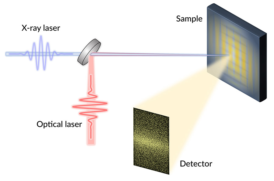
The team aimed infrared laser pulses at the YBCO sample to switch off its superconducting state, then used X-ray laser pulses to illuminate the sample and examined the X-ray light scattered from it. Their results revealed that regions of superconductivity and charge density waves were arranged in unexpected ways. (Courtesy Giacomo Coslovich/SLAC National Accelerator Laboratory)
Room-temperature superconductors could transform everything from electrical grids to particle accelerators to computers – but before they can be realized, researchers need to better understand how existing high-temperature superconductors work.
Now, researchers from the Department of Energy’s SLAC National Accelerator Laboratory, the University of British Columbia, Yale University and others have taken a step in that direction by studying the fast dynamics of a material called yttrium barium copper oxide, or YBCO.
The team reports May 20 in Science that YBCO’s superconductivity is intertwined in unexpected ways with another phenomenon known as charge density waves (CDWs), or ripples in the density of electrons in the material. As the researchers expected, CDWs get stronger when they turned off YBCO’s superconductivity. However, they were surprised to find the CDWs also suddenly became more spatially organized, suggesting superconductivity somehow fundamentally shapes the form of the CDWs at the nanoscale.
“A big part of what we don’t know is the relationship between charge density waves and superconductivity,” said Giacomo Coslovich, a staff scientist at the Department of Energy’s SLAC National Accelerator Laboratory, who led the study. “As one of the cleanest high-temperature superconductors that can be grown, YBCO offers us the opportunity to understand this physics in a very direct way, minimizing the effects of disorder.”
He added, “If we can better understand these materials, we can make new superconductors that work at higher temperatures, enabling many more applications and potentially addressing a lot of societal challenges – from climate change to energy efficiency to availability of fresh water.”
Observing fast dynamics
The researchers studied YBCO’s dynamics at SLAC’s Linac Coherent Light Source (LCLS) X-ray laser. They switched off superconductivity in the YBCO samples with infrared laser pulses, and then bounced X-ray pulses off those samples. For each shot of X-rays, the team pieced together a kind of snapshot of the CDWs’ electron ripples. By pasting those together, they recreated the CDWs rapid evolution.
“We did these experiments at the LCLS because we needed ultrashort pulses of X-rays, which can be made at very few places in the world. And we also needed soft X-rays, which have longer wavelengths than typical X-rays, to directly detect the CDWs,” said staff scientist and study co-author Joshua Turner, who is also a researcher at the Stanford Institute for Materials and Energy Sciences. “Plus, the people at LCLS are really great to work with.”
These LCLS runs generated terabytes of data, a challenge for processing. “Using many hours of supercomputing time, LCLS beamline scientists binned our huge amounts of data into a more manageable form so our algorithms could extract the feature characteristics,” said MengXing (Ketty) Na, a University of British Columbia graduate student and co-author on the project.
The team found that charge density waves within the YBCO samples became more correlated – that is, more electron ripples were periodic or spatially synchronized – after lasers switched off the superconductivity.
“Doubling the number of waves that are correlated with just a flash of light is quite remarkable, because light typically would produce the opposite effect. We can use light to completely disorder the charge density waves if we push too hard,” Coslovich said.
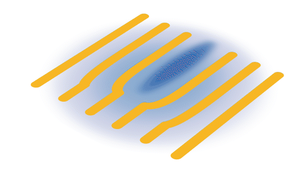
Blue areas are superconducting regions, and yellow areas represent charge density waves. After a laser pulse (red), the superconducting regions are rapidly turned off and the charge density waves react by rearranging their pattern, becoming more orderly and coherent. (Greg Stewart/SLAC National Accelerator Laboratory)
To explain these experimental observations, the researchers then modeled how regions of CDWs and superconductivity ought to interact given a variety of underlying assumptions about how YBCO works. For example, their initial model assumed that a uniform region of superconductivity when shut off with light would become a uniform CDW region – but of course that didn’t agree with their results.
“The model that best fits our data so far indicates that superconductivity is acting like a defect within a pattern of the waves. This suggests that superconductivity and charge density waves like to be arranged in a very specific, nanoscopic way,” explained Coslovich. “They are intertwined orders at the length scale of the waves themselves.”
Illuminating the future
Coslovich said that being able to turn superconductivity off with light pulses was a significant advance, enabling observations on the time scale of less than a trillionth of a second, with major advantages over previous approaches.
“When you use other methods, like applying a high magnetic field, you have to wait a long time before making measurements, so CDWs rearrange around disorder and other phenomena can take place in the sample,” he said. “Using light allowed us to show this is an intrinsic effect, a real connection between superconductivity and charge density waves.”
The research team is excited to expand on this pivotal work, Turner said. First, they want to study how the CDWs become more organized when the superconductivity is shut off with light. They are also planning to tune the laser’s wavelength or polarization in future LCLS experiments in hopes of also using light to enhance, instead of quench, the superconducting state, so they could readily turn the superconducting state off and on.
“There is an overall interest in trying to do this with pulses of light on very fast timescales, because that can potentially lead to the development of superconducting, light-controlled devices for the new generation of electronics and computing,” said Coslovich. “Ultimately, this work can also help guide people who are trying to build room-temperature superconductors.”
This research is part of a collaboration between researchers from LCLS, SLAC’s Stanford Synchrotron Radiation Lightsource (SSRL), UBC, Yale University, the Institut National de la Recherche Scientifique in Canada, North Carolina State University, Universita CAattolica di Brescia and other institutions. This work was funded in part by the DOE Office of Science. LCLS and SSRL are DOE Office of Science user facilities.
The number of cancer patients receiving proton beam therapy (PBT) – a newer, more targeted form of radiation therapy – is increasing, but Black patients are less likely to get this treatment than white patients, according to two recent studies published in JAMA Network Open.
Maria Vega, a member of Montana’s Fort Peck Assiniboine and Sioux Tribes, was jailed in 2015 after a suicide attempt. She is now part of a group of tribal members, academics, and policy experts proposing alternatives to the tribal policy of treating suicide as a crime. Photo by Sara Reardon / Kaiser Health News
The gap between suicide rates in rural and urban areas has grown, in part due to limited access to mental health services and privacy concerns in rural settings. Read more in my article in the American Journal of Nursing.
Did you make a New Year’s resolution to exercise more? And perhaps the more important question: Will you stick to your goal?
These questions are especially important for older adults, who are at a higher risk for chronic diseases such as dementia, cardiovascular disease, depression and anxiety. Physical activity can help reduce the risk for many of these conditions.
“We need to start thinking about these diseases [as diseases] of neglect, not necessarily of aging, that occur because people have not been able to maintain a lifelong pattern of healthy behavior,” said Randall Stafford, MD, PhD, a professor of medicine, in an article originally reported by Stanford’s BeWell.
Evolving intensity
Stafford explained that the exercises appropriate for any one person will likely evolve over his or her lifetime, but increasing physical activity at any age can quickly improve health.
Take my 92-year old relative Al, for instance. He started training and running marathons when he turned 40. In his 80s, he stopped running based on his doctor’s advice but kept hiking. These days, he walks a mile or rides his exercise bike for 30 minutes at a slow pace with breaks, along with strength and training exercises. His goal: Live an active, independent life.
But even if you’re not like Al (yet), it’s not too late; exercise doesn’t have to be something as intense as running a marathon.
“Even incorporating a few minutes of walking into one’s daily routine can be quite beneficial,” said Stafford. “Physical activity has benefits that are immediate as well as sustained.” And people often become better or more comfortable doing physical activities with practice, he said.
Expanding your mindset
Stafford’s other good news? You don’t have to do vigorous, gym-based exercises; joyful movements like gardening or dancing count. You’ll also get an extra social benefit if you share these physical activities with friends or family members, plus you are more likely to stick with the healthy behavior if you do it with others.
Stafford, however, stressed the importance of including strength training, core exercises and stretching — especially for people over 40 — to reduce muscle loss, maintain balance and stay flexible.
Finally, Stafford advised not to beat yourself up if you slide back into sedentary habits. Setbacks happen. Just try to get back into a routine as soon as you’re able.
This is a reposting of my Scope blog story, courtesy of Stanford School of Medicine.
An animation shows how charging and discharging a lithium battery test cell causes an island of “dead,” or detached, lithium metal to creep back and forth between the electrodes. The movement of lithium ions back and forth through the electrolyte creates areas of negative (blue) and positive (red) charge at the ends of the island, which swap places as the battery charges and discharges. Lithium metal accumulates at the negative end of the island and dissolves at the positive end; this continual growth and dissolution causes the back-and-forth movement seen here. SLAC and Stanford researchers discovered that adding a brief, high-current discharging step right after charging the battery nudges the island to grow in the direction of the anode, or negative electrode. Reconnecting with the anode brings the island’s dead lithium back to life and increases the battery’s lifetime by nearly 30%. (Greg Stewart/SLAC National Accelerator Laboratory.)
Menlo Park, Calif. — Researchers at the Department of Energy’s SLAC National Accelerator Laboratory and Stanford University may have found a way to revitalize rechargeable lithium batteries, potentially boosting the range of electric vehicles and battery life in next-gen electronic devices.
As lithium batteries cycle, they accumulate little islands of inactive lithium that are cut off from the electrodes, decreasing the battery’s capacity to store charge. But the research team discovered that they could make this “dead” lithium creep like a worm toward one of the electrodes until it reconnects, partially reversing the unwanted process.
Adding this extra step slowed the degradation of their test battery and increased its lifetime by nearly 30%.
“We are now exploring the potential recovery of lost capacity in lithium-ion batteries using an extremely fast discharging step,” said Stanford postdoctoral fellow Fang Liu, the lead author of a study published Dec. 22 in Nature.
Lost connection
A great deal of research is looking for ways to make rechargeable batteries with lighter weight, longer lifetimes, improved safety, and faster charging speeds than the lithium-ion technology currently used in cellphones, laptops and electric vehicles. A particular focus is on developing lithium-metal batteries, which could store more energy per volume or weight. For example, in electric cars, these next-generation batteries could increase the mileage per charge and possibly take up less trunk space.
Both battery types use positively charged lithium ions that shuttle back and forth between the electrodes. Over time, some of the metallic lithium becomes electrochemically inactive, forming isolated islands of lithium that no longer connect with the electrodes. This results in a loss of capacity and is a particular problem for lithium-metal technology and for the fast charging of lithium-ion batteries.
However, in the new study, the researchers demonstrated that they could mobilize and recover the isolated lithium to extend battery life.
“I always thought of isolated lithium as bad, since it causes batteries to decay and even catch on fire,” said Yi Cui, a professor at Stanford and SLAC and investigator with the Stanford Institute for Materials and Energy Research (SIMES) who led the research. “But we have discovered how to electrically reconnect this ‘dead’ lithium with the negative electrode to reactivate it.”
Creeping, not dead
The idea for the study was born when Cui speculated that applying a voltage to a battery’s cathode and anode could make an isolated island of lithium physically move between the electrodes – a process his team has now confirmed with their experiments.
The scientists fabricated an optical cell with a lithium-nickel-manganese-cobalt-oxide (NMC) cathode, a lithium anode and an isolated lithium island in between. This test device allowed them to track in real time what happens inside a battery when in use.
They discovered that the isolated lithium island wasn’t “dead” at all but responded to battery operations. When charging the cell, the island slowly moved towards the cathode; when discharging, it crept in the opposite direction.
“It’s like a very slow worm that inches its head forward and pulls its tail in to move nanometer by nanometer,” Cui said. “In this case, it transports by dissolving away on one end and depositing material to the other end. If we can keep the lithium worm moving, it will eventually touch the anode and reestablish the electrical connection.”
Boosting lifetime
The results, which the scientists validated with other test batteries and through computer simulations, also demonstrate how isolated lithium could be recovered in a real battery by modifying the charging protocol.
“We found that we can move the detached lithium toward the anode during discharging, and these motions are faster under higher currents,” said Liu. “So we added a fast, high-current discharging step right after the battery charges, which moved the isolated lithium far enough to reconnect it with the anode. This reactivates the lithium so it can participate in the life of the battery.”
She added, “Our findings also have wide implications for the design and development of more robust lithium-metal batteries.”
This work was funded by the DOE Office of Energy Efficiency and Renewable Energy, Office of Vehicle Technologies under the Battery Materials Research (BMR), Battery 500 Consortium and eXtreme Fast Charge Cell Evaluation of Li-ion batteries (XCEL) programs.
This is a reposting of a press release, courtesy of SLAC National Accelerator Laboratory.