
Tuberculosis was one of the deadliest known diseases, until antibiotics were discovered and used to dramatically reduce its incidence throughout the world. Unfortunately, before the infectious disease could be eradicated, drug-resistant forms emerged as a major public health threat — one quarter of the world’s population is currently infected with TB and 600,000 people develop drug-resistant TB annually.
New research at SLAC National Accelerator Laboratory is seeking to better understand how this antibiotic resistance develops, as recently reported in BMC Biology.
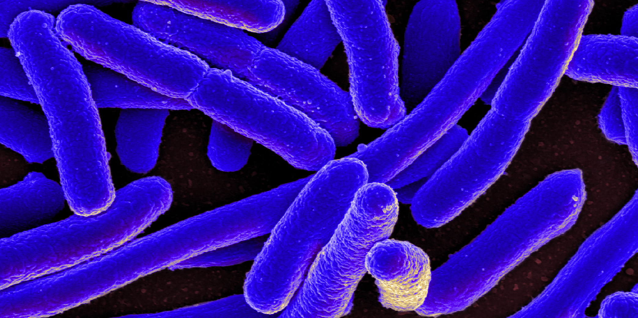
TB is caused by Mycobacterium tuberculosis bacteria, which attack the lungs and then spread to other parts of the body. The bacteria are transmitted to other people through the air, when an infected person speaks, coughs or sneezes.
These bacteria survive antimicrobial drugs by mutating. Their resilience is enhanced by the lengthy and complex nature of standard treatment, which requires patients to take four drugs every day for six to nine months. Patients often don’t complete this full course of TB treatment, causing the bacteria to evolve to survive the antibiotics.
Now, a team of international researchers has investigated an enzyme, called beta-lactamase, that is produced by the Mycobacterium tuberculosis bacteria. They wanted to understand the critical role this enzyme plays in TB drug resistance.
Specifically, the researchers made tiny crystals of beta-lactamase and mixed them with the antibiotic ceftriaxone. A fraction of a second later, they hit the enzyme-antibiotic mixture with ultrafast, intense X-ray pulses from SLAC’s Linac Coherent Light Source — taking millions of X-ray snapshots of the chemical reaction in real time for two seconds.
Putting these snapshots together, the researchers mapped out the 3D structure of the antibiotic as it interacted with the enzyme. They watched the bacterial enzyme bind to the antibiotic and then break open one of its key chemical bonds, making the antibiotic ineffective.
“For structural biologists, this is how we learn exactly how biology functions,” said Mark Hunter, PhD, staff scientist at SLAC and co-author on the study, in a recent news release. “We decipher a molecule’s structure at a certain point in time, and it gives us a better idea of how the molecule works.”
The research team plans to use their method to study additional antibiotics, observing in real time the rapid molecular processes that occur as the bacteria’s enzymes breakdown the drugs. Ultimately, they hope this knowledge can be used to design better antibiotics that can fight off these attacks.
This is a reposting of my Scope blog story, courtesy of Stanford School of Medicine.

.jpg)