Stanford Identifies Drug that May Improve Cardiac Stents

Stent in human coronary artery. (Wikimedia, Blausen gallery 2014)

Researchers from Stanford University School of Medicine believe they’ve found a drug for cardiac stents that can more effectively prevent stent complications.

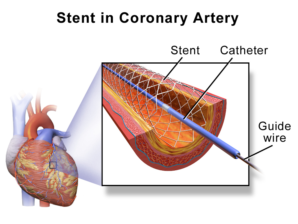
Over a million people in the U.S. each year undergo angioplasty heart surgery using a drug-coated stent to treat blocked arteries, according to the American Heart Association. A stent is a tiny wire mesh tube that is permanently implanted into the artery at the blockage point, creating a scaffold that props open the artery to reduce the chance of a heart attack. However, placement of bare metal stents can themselves damage the artery lining, causing scar tissue to grow and narrow the artery. Known as in-stent stenosis, this typically occurs 3-6 months after the surgical procedure and can lead to chest pain and even heart attacks.

To help prevent in-stent stenosis, doctors use stents coated with drugs that inhibit tissue regrowth to help prevent the blood vessels from reclosing. Unfortunately, these drugs can also inhibit beneficial regrowth of the vessel’s blood lining (endothelium) that aids the healing process. So patients still need to take blood-thinners for up to a year to reduce the risk of a blood clot developing in the stent and blocking the artery. This need for blood thinners is a serious problem for many people with other health issues; for instance, it means they can’t have surgery while taking the medication.

Stanford researchers have now identified a drug to coat cardiac stents that helps prevent in-stent stenosis without affecting the healing of the blood vessel lining. Their new research is described in a paper published this month in the Journal of Clinical Investigation. Dr. Euan Ashley, associate professor of cardiovascular medicine and genetics at Stanford University Medical Center, led the research team.

The researchers first sought to more fully understand the genetic pathways of coronary artery disease using a “big data” computational biology approach. Using data from previous studies, they analyzed large datasets of coronary artery tissue samples and genome information from patients who had developed in-stent stenosis after undergoing angioplasty and stenting. Based on network analyses, the researchers hypothesized that there is an increased risk of in-stent stenosis due to the interplay of two genes, GPX1 and ROS1.

GPX1 deficiency is known to be independently associated with coronary artery disease in humans. However, ROS1 expression is mostly known for its role in highly malignant cancers, such as lung cancers.

“We didn’t know anything about ROS1,” said Ashley in a press release. “It hadn’t been studied in cardiovascular disease. We knew it was an important gene in cancer. We thought, that’s odd, since the growth caused by stents is almost like a tumor.”

They confirmed their theory by performing an extensive series of laboratory experiments using human tissue samples and genetically engineered knockout mice. Some of these studies involved surgically implanting drug-coated stents in mice with clogged arteries. The researchers inhibited the ROS1 genes by coating these stents with crizotinib – a chemotherapy drug used to treat certain ROS1-positive lung cancers. They found that crizotinib inhibited in-stent stenosis without affecting the lining of the blood vessels.

“The major finding of the study is that artery stent disease acts surprisingly like a tumor in the blood vessel wall,” said Ashley in the press release. “Inhibiting it with nonspecific pharmaceutical agents, as we do now, leads to heart attacks from clots caused by lack of endothelial lining on the stent. Whereas, targeting it with the drug we use here, crizotinib, acts much more specifically and inhibits the disease without affecting the endothelium.”

A tiny mouse-sized stent used by Stanford researchers in their mice studies. (Courtesy of Euan Ashley)

Stanford researchers still have a lot more work to do before crizotinib-coated stents will be clinically available. However, this research should translate to the clinic more quickly since crizotinib is already an FDA approved drug.

This is a repost of my KQED Science blog.

New Imaging Method to Detect Heart Attack Risk

Image courtesy of NIH / Wikimedia Commons

785,000 people have an initial heart attack and another 470,000 people have a recurrent heart attack every year in the United States, according to the American Heart Association. This means that an American has a heart attack every 34 seconds and one dies from heart disease every minute. A new imaging technique may help identify who is at high risk.

The primary cause of heart attacks is clogged arteries. Arteries are blood vessels that carry oxygen-rich blood throughout the body. Blood flows easily in healthy arteries with smooth walls. But blood flow is reduced or blocked completely in clogged arteries, when a substance called plaque builds up on the inner walls of the arteries.

Artery-clogging plaque is made up of fat, calcium, cholesterol and other substances found in the blood. Over time, this plaque can harden and rupture. If it breaks apart, a blood clot can form on its surface and completely block the artery, preventing blood from reaching the heart muscle and causing a heart attack. If the blood flow isn’t quickly restored, the portion of the heart fed by the artery begins to die.

Coronary angiography is the “gold standard” way to identify these plaque blockages in the heart, but it’s an invasive surgical procedure. During a coronary angiography, a thin flexible tube called a catheter is put into a blood vessel in your arm, groin or neck and threaded into your coronary arteries. Then a special die is released through the tube, making your coronary arteries visible on X-rays pictures taken as the die flows through them.

New study results, recently published in The Lancet medical journal, show that these high-risk plaque blockages can also be identified using a non-invasive imaging technique. The study was carried out by Dr. Nik Joshi and his research team from the University of Edinburgh, the Royal Infirmary of Edinburgh and the University of Cambridge.

The study involved 40 people who had recently suffered a heart attack and 40 additional people who had stable chest pain (angina). The patients were given a standard coronary angiography and a non-invasive imaging PET-CT scan.

A PET-CT scan measures metabolic activity using positron emission tomography (PET) and anatomical structure using X-ray computed tomography (CT). A trace amount of radioactive drug is injected into the patient’s vein and used to produce 3D images. Joshi and his research team used a radioactive drug called sodium fluoride (NaF).

The study aimed to show how well a PET-CT scan using sodium fluoride detected plaques that had already ruptured or were at high risk of rupturing. The coronary angiography was used as a gold standard to identify the culprit plaque deposits that blocked the arteries.

The researchers measured the sodium fluoride distribution to determine if the artery-clogging plaques took up a significant amount of the drug. In 93% (37/40) of the people who had had a heart attack, significant sodium fluoride uptake was seen in the plaque responsible for the heart attack. The average drug uptake in these culprit plaque deposits was 34% higher than anywhere else in the heart.

In 45% (18/40) of the people with stable chest pain, culprit plaque deposits also took up significant amounts of the sodium fluoride drug. For both sets of patients, the culprit plaque deposits identified by PET-CT imaging were confirmed by histology or intravascular ultrasound to have high-risk characteristics such as calcification and a dead tissue core.

Further research studies with a broad range of patients are now needed before PET-CT sodium fluoride imaging is accepted as a standard clinical technique. These studies are likely to take several years to complete. If they confirm the initial promising results, the technique could then move immediately into clinics since it is already approved and commonly used for other applications.

“If the results are confirmatory then this technique has the potential to fundamentally alter the way we treat coronary artery disease,” concluded the investigators. “It could, for example, permit the identification of the vulnerable patient with single or multiple high-risk or silently ruptured plaques, providing an opportunity to treat and modify their risk to prevent future adverse cardiovascular events.”

This is a repost of my KQED Science blog.

%d bloggers like this: