The U.S. Preventative Services Task Force now recommends that many adults take a low to moderate dose of statin to reduce their risk of a heart attack or stroke, even if they don’t have a history of cardiovascular disease.
Statins are drugs that reduce the production of cholesterol by the liver — lowering bad cholesterol and triglycerides and raising good cholesterol. The task force comprehensively reviewed the literature on clinical trials and observational studies involving statin use. It concluded that the benefits of using statins outweighed the harms in some patients with increased risk of cardiovascular disease.
Douglas Owen, MD, a Stanford professor of medicine and director of the Center for Health Policy, was a member of the task force when the guidelines were developed. He summarizes the new recommendations in a recent news story:
“The task force recommends that clinicians offer statins to adults who are 40 to 75 years old and have at least one existing cardiovascular disease risk, such as diabetes, hypertension, high cholesterol or smoking. They also must have a calculated risk of 10 percent or more that they will experience a heart attack or stroke in the next decade. The task force recommends that clinicians use the American College of Cardiology/American Heart Association risk calculator to estimate cardiovascular risk because it provides gender- and race-specific estimates of heart disease and stroke.”
The task force hope these new recommendations will help clinicians better identify cardiovascular risk, so their patients can take steps to reduce their risk, such as eating a healthy diet, exercising and potentially taking a statin.
This is a reposting of my Scope blog story, courtesy of Stanford School of Medicine.
Forget fact checkers or polygraph tests. A functional magnetic resonance imaging (fMRI) brain scan might be the best way to tell if someone is lying.
According to a study from the University of Pennsylvania, our brains are more likely to give us away when we’re lying than sweaty palms, rapid breathing or spikes in blood pressure, the factors tracked by polygraph tests.
The researchers directly compared the ability of two techniques — fMRI and polygraph tests — to detect concealed information. They had 28 participants secretly write down a number between 3 and 8 on a slip of paper. Each participant then had both lie-detection tests, in random order, a few hours apart. During both sessions, they always answered “no” when asked if they had picked a certain number, which meant that one out of the six answers was a lie.
Three fMRI experts and three professional polygraph examiners then independently analyzed the results. The fMRI experts were 24 percent more likely to detect the lie than the polygraph experts, as recently reported in the Journal of Clinical Psychiatry.
Although the study wasn’t designed to evaluate the combined use of both techniques, the polygraph and fMRI results agreed correctly on the concealed number for 17 participants. So they plan to investigate in the future whether these techniques are complementary.
The study includes only a small number of participants, but the research team is encouraged by the results. “While the jury remains out on whether fMRI will ever become a forensic tool, these data certainly justify further investigation of its potential,” said Daniel Langleben, MD, first author and a professor in psychiatry, in a recent news release.
This is a reposting of my Scope blog story, courtesy of Stanford School of Medicine.
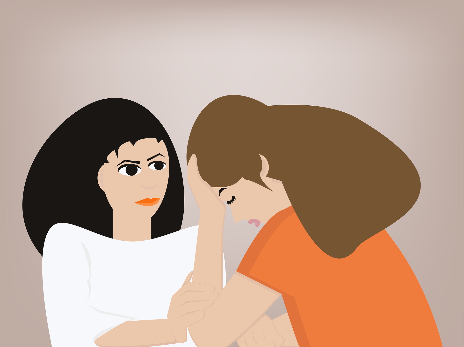
Most of us have no desire to think, or talk, about death. However, if we never talk about it, we leave our health providers and families guessing about how and where we want to die.
That’s why it’s important to communicate end-of-life preferences early, Philip Pizzo, MD, founder and director of the Stanford Distinguished Careers Institute, argues in a recent perspective in the Proceedings of the National Academy of Sciences.
It’s a topic he’s quite familiar with: Pizzo co-authored the Institute of Medicine’s report “Dying in America,” which addressed how to improve the quality of care for patients with advanced illnesses, without exacerbating the high costs of health care. In the perspective, he summarizes the IOM report’s key recommendations and provides an update.
Pizzo discussed the piece in an email:
“Unless we are facing an illness or event that makes the prospect of death imminent, most of us do not even think about the inevitability of our mortality. These conversations get slotted to times when death is more imminent and when our crisis-oriented decisions may not reflect our true preferences. That is why the IOM report recommended that conversations about death take place with our health care providers and families throughout life. Our thoughts and preferences about dying will vary at different stages of life and wellbeing.”
In the perspective, Pizzo describes the progress that has been made since the report was published. For instance, the Centers for Medicare & Medicaid Services in January 2016 began paying doctors to have end-of-life conversations with patients — a move Pizzo lauds as a major step.
Another important achievement, according to Pizzo, is the national stakeholder conferences that are now bringing constituencies together to implement the report’s recommendations.
He added:
“We witnessed before the IOM Committee began its work how rapidly public opinion can be swayed by political rhetoric. Thankfully since then, the public’s willingness to engage in conversations about death and dying have become better realized and books, like Atul Gawande’s Being Mortal or Paul Kalaniti’s When Breath Becomes Air, have helped to foster more enlightened conversations about dying.”
This is a reposting of my Scope blog story, courtesy of Stanford School of Medicine.
Many people, like me, have helplessly watched a loved one suffer and die from dementia. Now there is something you can do to help accelerate Alzheimer’s research — play a game.
The game, called Stall Catchers, is part of the EyesOnALZ project that uses citizen scientists to analyze Alzheimer’s research data. The game was developed by the Human Computation Institute, in collaboration with scientists from Cornell University, MIT and the University of California, Berkeley. The research team is trying to understand the association between reduced blood flow in the brain and Alzheimer’s disease.
The game features movies of real blood vessels in live mouse brains. Players must search for clogged vessels where blood flow is blocked, or stalled. Each movie is seen by many citizen scientists and then checked by a research scientist in order to quickly and accurately identify the stalls.
Past research has shown that Alzheimer’s is associated with the accumulation of beta amyloid proteins that clump together into sticky, neurotoxic aggregates called amyloid plaques. These proteins are normally cleared by the blood stream, but the formation of amyloid plaques slows down this clearance process.
Recent animal studies, performed by the Schaffer-Nishimura Lab at Cornell, suggest that improving blood flow in the brain may help reduce the devastating effects of amyloid accumulation. The researchers discovered that up to two percent of capillaries in the brains of Alzhiemer’s-affected mice were clogged — 10 times more than usual — and this caused up to a 30 percent decrease in overall blood flow in the brain.
“Advanced optical techniques have allowed us to peer into the brain of mice affected by Alzheimer’s disease,” said Chris Schaffer, PhD, the principal investigator in the Schaffer-Nishimura Lab, in a recent news release. “For the first time, we were able to identify the mechanism that is responsible for the significant blood flow reduction in Alzheimer’s, and were even able to reverse some of the cognitive symptoms typical to the disease.”
Now the main challenge for the Cornell researchers is the time-consuming process of manually analyzing all the brain movies to identify the stalled vessels. They need to study up to a thousand vessels for each animal. That’s why they collaborated with the experienced citizen teams at UC Berkeley and MIT to create the Stall Catchers game to get help from the public.
“Today, we have a handful of lab experts putting their eyes on the research data,” said Pietro Michelucci, PhD, the EyesOnALZ principal investigator, in a news story. “If we can enlist thousands of people to do that same analysis by playing an online game, then we have created a huge force multiplier in our fight against this dreadful disease.”
This is a reposting of my Scope blog story, courtesy of Stanford School of Medicine.
Millions of patients suffering from fibromyalgia often experience widespread musculoskeletal pain, sleep disturbances, fatigue, headaches and mood disorders. Many also struggle to even get diagnosed, since there are currently no laboratory tests for fibromyalgia and the main symptoms overlap with many other conditions. However, new research may help.
Scientists from the University of Colorado, Boulder may have found a pattern of brain activity that identifies the disease. They used functional MRI (fMRI) scans to study the brain activity of 37 fibromyalgia patients and 35 matched healthy controls, while the participants were exposed to a series of painful and non-painful sensations.
As reported recently in the journal PAIN, the research team identified three specific neurological patterns correlated with fibromyalgia patients’ hypersensitivity to pain.
Using the combination of all three patterns, they were able to correctly classify the fibromyalgia patients and the controls with 92 percent sensitivity and 94 percent specificity — meaning that their test accurately identified 92 percent of those with and 94 percent of those without the disease.
Tor Wager, PhD, senior author and director of the school’s Cognitive and Affective Control Laboratory, explained the significance of the work in a recent news release:
“Though many pain specialists have established clinical procedures for diagnosing fibromyalgia, the clinical label does not explain what is happening neurologically and it does not reflect the full individuality of patients’ suffering. The potential for brain measures like the ones we developed here is that they can tell us something about the particular brain abnormalities that drive an individual’s suffering. That can help us both recognize fibromyalgia for what it is – a disorder of the central nervous system – and treat it more effectively.”
More research is needed, but this study sheds a bit of light on this “invisible” disease.
This is a reposting of my Scope blog story, courtesy of Stanford School of Medicine.
Have you tried to talk to a friend or family member about cancer? It’s not easy. You might have blurted out something offensive, offered advice you weren’t quite sure about, or tried to minimize cancer’s severity or prevalence. Or maybe you just avoided the conversation entirely.
Even those with medical training struggle with cancer discussions. In response, a London-based nonprofit has created a free online course called “Talking About Cancer,” which offers strategies for discussing cancer risks, preventing and screening.
The three-hour course — which is designed for health-care workers, counselors, volunteers and others — is organized into short, self-paced modules made up of videos, quizzes, online discussions and role-playing with actors. I recently spoke with one of the course organizers, former journalist and Stanford alumni David Risser.
What is the course like?
“The course quickly reviews cancer myths and facts, and then concentrates on how to have confident conversations about cancer prevention. It’s an important area, because more than four in ten cancer cases could be prevented by lifestyle changes. Another goal is to boost early diagnosis, by training people to encourage others to see a doctor.
We wanted to present an engaging course with varied activities, including a ‘real-life’ narrative that runs through the course. We hired improv actors to play two characters, Anita and Brian. Anita has health difficulties but is reluctant to see a doctor. Brian feels invincible, but has multiple habits — smoking, drinking too much, a poor diet, and even refusing to use sunscreen — that put him at higher risk… We were amazed at how strongly participants identified with the experiences of these characters, leading to passionate discussions about effective ways to talk about cancer.
The course is led by our cancer awareness trainers — two nurses with experience in engaging doctors, nurses and people without medical training. It’s open now, but you must sign up by October 31. Otherwise, we plan to offer another free, public run of the course early next year.”
What inspired you to get involved?
“I am a Stanford graduate with a B.A. in history, and a past managing editor of The Stanford Daily. I joined Cancer Research UK after a 28-year career in journalism, in part because of my own experience with several family members who have had cancer. I struggled to talk to one family member who didn’t seem to want to talk about her cancer. That made it easy for me to avoid talking about it, which wasn’t the ideal outcome.
It was also difficult to talk to another family member about being more proactive about her medical care, which was inadequate at the start. It’s still difficult to know whether or how to talk to friends about prevention.”
What have you learned while working on this project?
“I learned that there are widespread gaps in knowledge about cancer, from causes to treatments to chances of survival. For example, many people don’t know that obesity is the second most-preventable cause of cancer or that alcohol is linked to a range of cancers.
I think the most common mistake when talking about cancer is feeling you have to know everything. It’s more effective to say ‘I don’t know, how do you think we can find out?’ or `What have you thought about doing?’ than to avoid the subject, make pronouncements or communicate incorrect information. Another common mistake is failing to listen, listen, listen, rather than fix everything. Sometimes it’s better to guide the person through what they are feeling and what they are concerned about, keeping it about them and not about you or your solutions. The bigger issue in both cases is to gently help people see what they can do that works for them.
While the course focuses on cancer information, prevention and early diagnosis, most of it is about how to have conversations about difficult subjects. The greatest lesson in the course is how to talk to people in ways that encourage behavior change. And that can save lives.”
This is a reposting of my Scope blog story, courtesy of Stanford School of Medicine.
It isn’t news that we need calcium to keep our bones dense and strong. If you don’t get enough calcium in your diet, or if your body doesn’t absorb enough, your bones can become brittle and fragile. But calcium is more than just an essential part of your bone structure. A new research study shows that it also plays a major role in regulating the cells that control bone formation.
Biochemist Michael Rape, PhD, and his colleagues at University of California, Berkeley studied how bone cells form in very early embryos. In particular, they investigated neural crest cells that help develop bones in the head and face. Without enough neural crest cells, an embryo dies or has a craniofacial disorder like Treacher Collins syndrome, which is characterized by deformed ears, eyes and cheekbones.
The Berkeley researchers weren’t expecting to discover a new role for calcium. Instead, they were investigating how to turn on enzymes like CUL3 to form normal, healthy bones. Previous research showed that CUL3 enzymes can trigger undifferentiated stem cells to become neural crest cells during embryo development. In this study, the team learned that calcium is essential to help CUL3 trigger proper bone growth — surprising results that were recently reported in Cell.
“Our research basically identifies calcium not only as a structural element of bone, which makes the bone strong and sturdy, but also as a signaling molecule for bone formation that we hadn’t appreciated before, which can be used to turn enzymes on and off. …. This means that you basically have many different steps that come together in order to form a bone, and that they are beautifully orchestrated by calcium.”
This research became more personal to Rape when he was visited by Francis Smith, PhD, a Treacher Collins syndrome patient and postdoctoral research fellow at the University of Colorado. Rape said in the video above, “When you talk to patients of a disease, it shows you the direct consequences of your work and the urgency of your work becomes much more apparent.”
Further research is needed before these results can be used to help trigger proper bone growth for patients, but the researchers are hopeful. “The more you understand about each of these steps, the easier it is to focus your applied research onto things that matter and change something for these patients,” Rape said in the news release.
This is a reposting of my Scope blog story, courtesy of Stanford School of Medicine.
Football season has begun, reviving concern and discussion over sports-related concussions.
The American Academy of Pediatrics defines a concussion as a direct hit to the head or jarring blow to the body that gets transmitted to the head, resulting in a rapid onset of short lived impairment of neurological function. However, some controversy surrounds even this definition. So I reached out to Jessica Little, PhD, director of clinical research and operations at the Stanford Concussion and Brain Performance Center, to learn more about concussion research and Stanford’s clinical study of teenage athletes.
What should we know about concussions?
“I think it is important to note that concussions are still not well understood. There are hundreds of different definitions of ‘what is a concussion’ and there is currently no single evidence-based consensus on how to identify and treat concussions.
Research has shown that one of the biggest risk factors for sustaining a concussion is a history of having a prior concussion. There is a ‘window of vulnerability’ — the concept that a person experiencing symptoms of concussion is more vulnerable to incurring a second concussion during this time, as the brain has not yet fully recovered. If a truly concussed athlete has problems paying attention or is not coordinated, they can then be vulnerable to another injury. Protocols are often used to track signs and symptoms of concussion, and athletes are not allowed to return to play until these have resolved. However, it would be helpful to have more precise ways to measure attention and coordination on the sidelines to keep impaired athletes out of contact sports until those skills recover.
The vast majority of people with a concussion recover fully after the injury, though not all symptoms may improve at the same rate and everyone recovers a little differently.”
Describe your clinical study for athletes between 12-17 years of age.
“Our study just closed recruitment and we’re prepping all the data for analysis, so it is an exciting time. The study was called EYE-TRAC Advance, short for Eye-Tracking Rapid Attention Computation. Our lab used a specific type of eye-tracking called ‘circular smooth pursuit’ where an athlete follows a dot that moves at predictable speed around a circle. The eye-tracking was in the form of custom-designed portable “goggles,” using built-in cameras and infrared pupil detection.
Our hypothesis is that people without a concussion can ‘sync-up’ with the way the dot is moving pretty easily, while a person with a concussion has a disruption in their ability to focus and pay attention. You often hear people saying that they feel “off” or “out of sync” following a concussion, and we’re trying to quantify that experience. For the study, we baseline tested athletes (before sports participation) with the eye-tracking, as well as other neurocognitive tests that measured things like attention and reaction time. If the athlete later got a concussion, we tested them again as soon as possible and again at 1, 3 and 12 months after the injury. In this way, we’re able to get a clear picture of how their brain recovered over time.
Overall, we reached out to over 60 different organizations and recruited over 1,400 people. We had a specially outfitted ‘mobile testing center’ RV. This allowed us to literally drive up to the side of an athletic field and perform the testing on-site at the school or organization, which really reduced common barriers to participating in a research study, such as the costs and time associated with transportation to and from appointments.”
Can technology play a significant role in preventing concussions?
“A lot of current technologies focus on diagnosing a concussion, but there are far fewer that actually focus on preventing concussions. There are some technologies that measure an athlete’s gait and vestibular-balance ability. If there are impairments, the athletes can be provided skill training to improve any deficits, thus reducing the risk of injury. Other technologies, such as helmet technologies, may be helpful in reducing the instance of skull fractures and other serious injuries, but they haven’t yet proved effective at preventing a concussion — that is caused more by brain rotation, which a helmet can’t fully protect against. One possible preventative solution could come from a neck device that stabilizes the rotational forces while still allowing neck movement at low accelerations, so athletes can move about freely until it senses a potentially dangerous level of force.”
Are there issues with under-reporting concussions?
“Historically, there have been some issues with individuals under-reporting symptoms that would lead to a diagnosis of a concussion. This is often motivated by the idea that they should ‘suck it up,’ ‘don’t want to let the team down’ or the fact that their ability to perform athletically is tied to keeping an athletic scholarship. There is research happening in the field right now trying to figure out the best way to dismantle these types of beliefs and make it more likely that athletes can be properly identified, given the treatment they need, and hopefully continue to safely engage in their sport.”
This is a reposting of my Scope blog story, courtesy of Stanford School of Medicine.
Medical students frequently turn to mnemonics to master human anatomy, but they’re usually just catchy phrases. Now, Nick Love, a second-year Stanford medical student, has created a more entertaining way to memorize anatomy: a set of illustrated mnemonics, which he has published in the form of a book and website. I recently spoke with Love about his project.
What inspired you to illustrate the anatomic mnemonics?
“When I began medical school, I was totally unaware as to the central role mnemonics play in medical education and beyond. They are everywhere! Their sometimes wacky and ridiculous wordings intrigued me — I wondered if they could serve as a unique source of ‘found imagery,’ starting points for visual exploration. I brought up this idea with Audrey Shafer, MD, director of the Biomedical Ethics and Humanities medical school track, and she kindly encouraged me and linked me up with an awesome mentor for the project, pediatric anesthesiologist and painter Samuel Rodriguez, MD.”
Where did you get the mnemonics and how did you choose your illustration style?
“They are all essentially common med school mnemonics. Fourteen of the 16 mnemonics were passed on to us as medical students, mainly by our clinical anatomy teaching assistants via the ‘whiteboards’ in the anatomy lab. I sourced one mnemonic directly from the internet, and I altered another because its original form was too raunchy for publication. At the moment, I am, unfortunately, too behind on too many things to add more.
In terms of illustration, I was motivated to try a digital-analog-digital process. I’m currently intrigued by combining the reproducibility of computer-aided illustration with the inherent chaos of spreading paint or ink. Also, I wanted to maximize color usage, insert a bit of whimsy into the illustrations and experiment with recursive imagery.”
Do you have a favorite mnemonic?
“My favorite mnemonic is ‘canned soup, really good in cans.’ It helps one remember the branches of the descending aorta — canned soup, really good in cans, representing celiac, superior mesenteric, renal, gonadal, inferior mesenteric, and common iliac arteries. The phrase ‘canned soup, really good in cans’ strikes me as rather humorous, like it was made for an ad campaign when soup was first put into cans. Genius, whoever came up with it.”
Do you have any art training? Who are your favorite artists?
“Before coming to medical school, my training was mainly in science. However, last year I took two art classes at Stanford, ‘Digital Photography’ and ‘Video Compositing,’ both of which were awesome. As a kid, I mostly played sports, video games and outside. The desire to make things came later.
Do you hope to include art somehow in your future medical practice?
“I’m very much interested in learning more about what is referred to as the ‘art of medicine,’ and I hope to have the time to keep creating. At the moment, I’m most drawn to visually-based medical specialties, such as dermatology, pathology, radiology and nuclear medicine.”
This is a reposting of my Scope blog story, courtesy of Stanford School of Medicine.
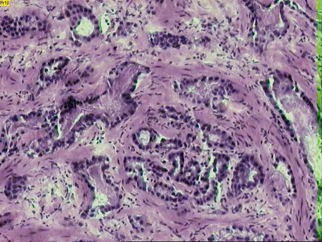
Many men with prostate cancer have slow-growing tumors that don’t require aggressive treatments such as surgery or radiation therapy, whereas others have rapidly-growing prostate tumors that are life threatening. Distinguishing between these patients with indolent versus aggressive disease is a major challenge, but researchers have just made a significant step towards identifying genetic risk factors for prostate cancer prognosis.
A multi-institutional research team has identified three distinct molecular subtypes of prostate cancer, which are correlated with survival rates and may guide future treatment planning. Their study results were presented at the annual meeting of the American Society of Radiation Oncology by Daniel Spratt, MD, an assistant professor in radiation oncology at the University of Michigan Heath System.
Spratt explained in a recent American Society of Radiation Oncology press release:
“Tumors that appear similar under a microscope can behave very differently, from a clinical standpoint. One promise of genomic analyses is to elucidate subtypes of cancer based on the genetics of the tumor rather than merely how they look or what size they are.”
The research team analyzed the RNA expression patterns of 4,236 primary prostate cancer samples taken from nine independent groups of men, who had their prostate surgically removed to treat primary prostate cancer. The investigators’ statistical clustering analysis identified three distinct patient groups based on 100 key genes, which they named the Prostate Cancer 100. These study results were then validated using samples from over 2100 patients.
The subtypes were found to be correlated with androgen receptor activity, ERG oncogene expression and other factors known to promote prostate tumor growth. They were also correlated with how long patients survived without metastasis. The distant metastasis-free survival rates varied among the three subgroups — 73.6 percent for group A, 64.4 percent for group B and 57.1 percent for group C — showing that subtype A patients had the most favorable prognosis.
Furthermore, the study found that subtype B and C patients responded significantly better to postoperative radiation therapy, which was used after the prostate was surgically removed in order to kill any remaining cancer cells. This is important because radiation therapy has many potential side effects, including impotence and incontinence.
Spratt summarized in the press release:
“We believe that these subtypes reflect truly distinctive underlying biology and that this work represents a significant advance in our understanding of prostate cancer biology. Moreover, our findings identify numerous genes and enriched biologically active pathways in prostate cancer that have been underappreciated to date but may be potential targets to improve cure rates in this disease by developing new targeted therapies.”
This is a reposting of my Scope blog story, courtesy of Stanford School of Medicine.


