Imagine you visit a doctor in a far-off land with a different language. Although you have an interpreter, the doctor barely looks at you — instead relaying all information through the interpreter. You feel extra, ignored. If anything, you are building a relationship with the interpreter, not the doctor.
And that’s not good, VJ Periyakoil, MD, clinical associate professor of medicine, points out in the video above.
Our goal is to talk with the patient, through the interpreter, not talkaboutthe patient,tothe interpreter.
The video is part of the Stanford Cross Cultural Medicine Microlecture Series, a series of very short talks (about one to five minutes) that aims to bridge the growing communication gap between doctors and their patients as the U.S. population becomes older and more diverse. There are already11 millionAmericans that are nonliterate in English and 25 million with only limited English proficiency.
Are doctors prepared?
These talks highlight key issues in cross-cultural encounters, including a range of practice tips for health professionals provided by experienced medical interpreters and from Periyakoil. The videos typically end with a take-home message listing the problem and solution — making it easy to quickly learn the concept. Periyakoil and her colleagues hope that health professionals will use the series as a tool to reflect on their own practice.
Microlecture 4emphasizes the importance of talking directly to the patient even when working with a medical interpreter. Patients with limited English proficiency have the right to complete healthcare information, as well as the right to the therapeutic bond between every doctor and patient.
There are currently 16 microlectures posted on thewebsite, but many more are on their way. A total of 44 microlectures have been made and two new ones are being released each week. The lectures also build off recommendations developed in a paper on ethnogeriatrics by the American Geriatrics Society.
This is a reposting of my Scope blog story, courtesy of Stanford School of Medicine.
Approximately 22,280 women will be diagnosed with ovarian cancer, and an estimated 14,240 will die from the disease in the United States this year. Ovarian cancer is deadly in part because the early warning signs are limited and nonspecific — such as abdominal bloating, pelvic pain, a frequent need to urinate and quickly feeling full when eating — so the symptoms are often blamed on other more common conditions. And pap smears don’t detect it, so only 15 percent of ovarian cancer cases are detected before the disease has spread to other tissues.
In 2009, the Department of Defense Ovarian Cancer Research Program created a unique, interactive virtual academy for early-career ovarian cancer researchers, called the OCRP Ovarian Cancer Academy. I recently spoke with a new member of the academy, Erinn Rankin, PhD, assistant professor of obstetrics and gynecology and radiation oncology, about the program and her research
What is unique about the Ovarian Cancer Academy?
The program is a really special funding mechanism that pairs early-career investigators with a local university mentor and a secondary mentor from another university. It provides early-investigators access to the ovarian cancer community, mentorship from established researchers and networking opportunities. Once a year, all members meet together in person. We also have monthly webinars that cover topics for early career development, such as introducing us to patient advocacy groups. And we discuss our individual research and try to identify areas where we can collaborate.
It’s a great grant that will support my research for five years with a total of about one million dollars. But really the access to all the mentors, investigators and networking is more important to my career than the money. I’m very honored and privileged to be part of it. I have a really supportive mentor, Jonathan Berek, MD, who is chair of obstetrics and gynecology here at Stanford. He’s fostered my career in ovarian cancer research. He’s also provided me with the opportunity to collaborate with other great researchers, like Oliver Dorigo, MD, PhD.
What are the major challenges of ovarian cancer diagnosis and treatment?
Ovarian cancer is a highly metastatic and deadly disease. Most patients are diagnosed with advanced metastatic disease. The standard of care for these patients is quite striking. They go through a surgical debulking and then they are treated with conventional chemotherapy. Unfortunately, most of them become resistant to the chemotherapy after multiple cycles, so they end up succumbing to their disease.
It’s such a devastating cancer. If we can make an impact for these patients, it would be wonderful. Since we don’t have good mechanisms to detect the disease earlier, a good way to improve overall survival is to develop new therapies that can effectively treat resistant metastatic disease and prevent recurrence.
What is the specific focus of your ovarian cancer research?
I study how the microenvironment of a tumor influences metastatic progression. In particular, I focus on how hypoxia, or low oxygen supply, drives ovarian cancer metastasis and its resistance to therapy. Virtually every solid tumor has areas of hypoxia. And hypoxia in these tumors is often associated with poor response to therapy, further metastatic progression and poor patient survival. This is the case in ovarian cancer.
We’ve identified a new, really exciting cancer therapy target — a cell receptor enzyme called axl that has a molecular link to hypoxia. Axl is highly expressed in ovarian cancer metastatic lesions in comparison to normal ovary tissue. We identified axl as a key factor regulating ovarian cancer metastasis through genetic means. We then collaborated with Amato Giaccia, PhD, in the radiation oncology department and Jennifer Cochran, PhD, in the bioengineering department to develop a novel therapeutic agent to target axl for metastatic therapy.
Our research now focuses on how our anti-axl therapy works to treat advanced metastatic ovarian cancer. We’re testing it in combination with the standard of care, which is chemotherapy, in two preclinical mouse models of ovarian cancer. We’re really excited. Our anti-axl therapy appears to be a highly potent and safe hypoxia inhibitor. Once we generate more preclinical data, we plan to take this agent into clinical trials. I hope our therapy will make a difference for these patients.
This is a reposting of my Scope medical blog story, courtesy of Stanford School of Medicine.
Dr. Marcus Maydew, radiologist from Creighton University, reviews an x-ray (Offutt Air Force Base).
As a Hodgkin’s lymphoma survivor, I’ve had plenty of CT scans, mammograms, chest X-rays and MRIs during my diagnosis, therapy and 20 years of follow-up care. So I’ve interacted with radiologists at many Bay Area clinics, including Stanford where I was treated — that is, if you count getting summary reports in the mail as “interactions.”
This type of interaction may be changing with the growing movement toward patient-centered care, which is a critical component of the new American College of Radiology’s Imaging 3.0 initiative. Some radiologists are now going beyond image interpretation by discussing test results directly with their patients.
“Many interventional radiologists are now creating their own clinics, seeing patients and following them like any other surgeon,” Sandip Biswal, MD, a Stanford associate professor of radiology, told me. “Patient interactions are also quite heavy in mammography, particularly if the radiologist sees something suspicious.”
As radiologists come out of their reading rooms, many need to improve their communication skills, and a new training program at UMass Memorial Medical Center, called “Coming Out of the Dark,” teaches first and fourth-year radiology residents effective communication skills through role-playing. The program is led by Carolynn DeBenedectis, MD, an assistant professor of radiology there.
The participants practice six scenarios, such as delivering bad news from breast imaging tests, with trained actors performing as the patients. They are evaluated by both the patient actors and attending radiologists with prior communication skills training. The sessions are also videotaped and reviewed with the residents. The same participants return two weeks later to role-play six similar scenarios in order to evaluate their improvement.
At the Radiological Society of North America 2015 meeting, DeBenedectis reported on last year’s pilot program results. Participants were graded using a standard communications assessment scale and their scores on average improved about 5 percent between the two sessions — from 74 percent to 79 percent for first-year residents. More importantly, participants found the training useful, as reported in a recent online story.
We could probably all benefit from improved communication skills. However, there is some controversy over whether diagnostic radiologists should discuss imaging results directly with their patients after their scans. Biswal explained to me:
In the patient’s best interest, we really need to take a team approach. The primary care physician or referring specialist has the best understanding of what the patient is going through, so they can better convey the news. For radiologists to sit down with a patient and give them imaging results without knowing their full story can be potentially dangerous. There is an art to conveying this type of information that takes years of practice. I think of it like this: if it was my mother, how would I want her to be treated?
This is a reposting of my Scope blog story, courtesy of Stanford School of Medicine.
A team of researchers co-led by David Relman, MD, professor of medicine and of microbiology and immunology, has discovered previously unknown species of bacteria in dolphins trained by the U.S. Navy.
You’ve probably heard of security dogs that help sniff out drugs, bombs or land mines — the U.S. Navy uses dolphins, the dogs’ marine equivalent, to protect ships and submarines by detecting sea mines and underwater intruders.
The researchers are cataloging the bacterial communities living inside the dolphins at the Navy’s Marine Mammal Program in San Diego. They analyzed samples from the dolphins’ mouths, stomachs, rectums and respiratory tracts. Their results were recently reported in Nature Communications.
The research team found a startling diversity of bacteria, especially from the dolphins’ mouths. “About three quarters of the bacterial species we found in the dolphins’ mouths are completely new to us,” Relman said in an online piece.
The researchers also tested the Navy’s sea lions and the surrounding seawater. The newly discovered bacteria found in the dolphins were not seen in the sea lions, even though the dolphins and sea lions were fed the same fish and swam in the same water. The bacteria in the seawater were also very different from the bacteria in the marine mammals.
Relman began working with the Navy 15 years ago to help keep the Navy dolphins healthy. However, their research may have a much wider impact, Relman explained in the story:
There’s a lot of concern about the changing conditions of the oceans and what the impact could be on the health of wild marine mammals. We would love to be able to develop a diagnostic test that would tell us when marine mammals are beginning to suffer from the ill effects of a change in their environment.
The research team plans to expand their study to include other marine mammals, including sea otters, harbor seals and elephant seals.
This is a reposting of my Scope blog story, courtesy of Stanford School of Medicine.
Opiates produce a sense of euphoria that is highly addictive. If addicts stop taking the drugs, they are faced with opiate withdrawal, which can feel like the worst imaginable stomach flu with symptoms that include muscle aches, sweating, nausea, vomiting, diarrhea and a runny nose.
Stanford researchers have identified and suppressed the neural pathway responsible for theses withdrawal symptoms in opiate-addicted mice, as reported in Nature.
Xiaoke Chen, PhD, the lead investigator and an assistant professor of biology, explains in a news release:
Most research that studies drug addiction is focused on the reward pathway because that is the reason you start to take drugs, but people who really get addicted also take drugs to get rid of the withdrawal effect. This is especially important in opiate addiction.
Chen’s team studied the nucleus accumbens, a group of neurons that plays a key role in addiction through its response to both rewarding and aversive stimuli. They used fluorescent proteins to identify a clear link between the nucleus accumben and another brain center associated with drug-seeking behavior called the paraventricular nucleus of the thalamus (PVT).
Next, the researchers used optogenetics to turn neurons in this nucleus accumben-PVT pathway off, by introducing light-sensitive molecules and then hitting them with light from an optical fiber. The news release explains:
Using optogenetic tools, the scientists were then able to revert the pathway to its original strength, effectively erasing the effects of the drug. Although the research was conducted in mice, Chen said that it suggests that reprogramming the circuit holds promise for treating opiate addiction in humans.
Chen’s research may guide the development of treatments for many people with exaggerated aversive response to stimuli, including those with drug addiction, anxiety and depression.
This is a reposting of my Scope blog story, courtesy of Stanford School of Medicine.
The levels of gadolinium in the San Francisco Bay have been steadily increasing over the past two decades, according to a study recently published in Environmental Science & Technology. Gadolinium is a rare-earth metal and the potential long-term effects of its exposure have not been studied in detail.
They found the gadolinium levels to be much higher in the southern end of the Bay, which is home to about 5 million people and densely populated with medical and industrial facilities, than in the central and northern regions. They also observed a sevenfold rise in gadolinium concentration in the South Bay over that time period.
The study attributes the rising level of gadolinium contamination largely to the growing number of magnetic resonance imaging (MRI) scans performed with a gadolinium contrast agent. A gadolinium contrast agent is used for about 30 percent of MRI scans to improve the clarity of the images. It is injected into the patient then excreted out of the body in urine within 24 hours.
Lewis Shin, MD, assistant professor of radiology and a MRI radiologist, explained to me the importance of using intravenous gadolinium contrast agents:
“Gadolinium contrast agents allow us to detect abnormalities that would otherwise be hidden from view and to improve our characterization of the abnormalities that we do find. Gadolinium is not always used; for example, if a physician is just concerned about identifying a herniated disk in the spine, an MRI without contrast agent is sufficient.
However, gadolinium is routinely administered to detect and characterize lesions if there is a clinical concern of cancer. Also, if a patient was previously treated for cancer, gadolinium administration is often extremely helpful to detect early recurrences. In summary, MRI with a gadolinium contrast agent greatly improves our ability to make an accurate diagnosis not only for cancer but for many other disease processes as well.”
According to UCSC researchers, gadolinium is not removed by standard wastewater treatment technologies, so it is discharged by wastewater treatment plants into surface waters that reach the Bay.
Shin expressed some surprise when he learned about this study:
“The majority of radiologists probably don’t even think about gadolinium once it’s excreted out of a patient’s body. Of course it’s concerning that there is a rise in gadolinium levels in the environment, but the next questions are how is this impacting the environment and whether there is a safe level or not? Since most of the gadolinium contrast agents used for MRI studies are excreted through the urine within 12 to 24 hours, one strategy to reduce environmental release of gadolinium could be to collect patients’ urine for a brief period of time for proper disposal or even recycling of the gadolinium itself.”
The UCSC researchers assert that the current levels of gadolinium observed in San Francisco Bay are well below the peak concentrations that could pose harmful effects on the aquatic ecosystem. However, they recommend in their paper, “new public policies and the development of more effective treatment technologies may be necessary to control sources and minimize future contamination.”
This is a reposting of my Scope blog story, courtesy of Stanford School of Medicine.
Many academic researchers are tenacious, spending years in the lab studying the processes that lead to human diseases in hopes of developing treatments. But they often underestimate how difficult it is to translate their successful discovery into a drug that will be used in the clinic.
That’s why Daria Mochly-Rosen, PhD, founded SPARK, a hands-on training program that helps scientists move their discoveries from bench to bedside. SPARK depends on a unique partnership between university and industry experts and executives to provide the necessary education and mentorship to her academic colleagues.
In recent years, Stanford’s program has sparked identical programs throughout the world; at TEDMED 2015, Mochly-Rosen described this globalization. I recently spoke with her about the SPARK Global program, which she co-directs with Kevin Grimes, MD, MBA.
How has SPARK inspired similar programs throughout the world?
We’ve found our solution for translational research to be particularly powerful. Of the 73 completed projects at Stanford, 60 percent entered clinical trials and/or were licensed by a company. That’s a very high accumulative success rate. So I think it has showed other groups that we have a formula that really works – a true partnership with academia and industry. It’s the combination of industry people coming every week to advise us and share lessons learned and our out-of-the-box, risk-taking academic ideas that makes SPARK so successful.
We feel that what we’ve learned is applicable to others. Kevin and I also feel very strongly that universities need to take responsibility to make sure inventions are benefitting patients. So we’re trying to do our part.
How do you and Dr. Grimes help develop the global programs?
When a university asks about our program, we invite them to come visit us for a couple of days so they can talk to SPARKees (SPARK participants), meet SPARK advisors and watch our weekly meeting. Sometimes they also ask Kevin and I to come to their country to help set up a big event or assist in other ways. If they begin a translational research program at their institution, we offer for them to be affiliated with SPARK Global. Everyone is invited.
There are now SPARK programs throughout the world, including the United States, Taiwan, Japan, Singapore, South Korea, Australia, Germany and Brazil. We are also working with other countries, including Norway, Israel, Netherlands, Poland and Finland to help them start a program.
Do researchers in other countries face the same challenges as those in the US when developing new drugs?
There are many common challenges. And there are also some advantages and challenges that are different in other places. So it’s a mix, both within and outside the US.
There are several key components to the success of translation research. It’s important to have a good idea. It’s even more important to have good advisors from industry to help develop the idea. And it’s very important that the people involved are open-minded and are not inhibited by hierarchical structures. In some places, there is a big problem with hierarchy – particularly in parts of Europe and East Asia. In some cultures, it’s also difficult to get experts to volunteer and academics can’t afford to pay multiple advisors. Also, some universities don’t have a good office of technology to help with patent licensing, which can be a major challenge.
You recent held the first International SPARK conference. Do you have future events planned?
The first international SPARK conference was held last summer in Taiwan. We only invited those with an existing SPARK program, because it was an organizational meeting. We spent a lot of time discussing what we want to do together.
The next SPARK Global meeting will be open to every university and will be held at Stanford this fall. There will be half a day for those thinking about starting a new SPARK program at their institution, and then one-and-a-half days for those already involved. We’ll celebrate SPARK’s 10-year anniversary and the formation of SPARK Global. Our overall agenda is to continue to promote SPARK-like programs in universities, as well as come up with ideas that the global network can work on together.
This is a reposting of my Scope blog story, courtesy of Stanford School of Medicine.
Remember all the rumors that you heard about sexuality and fertility as a teen (or even a 20-something or a 30-something)? It’s hard to sort out fact from fiction.
According to the Institute for Reproductive Health (IRH) at the Georgetown University Medical Center, an accurate understanding of sexuality and fertility is surprisingly low around the world. That’s why IRH has created an online quiz to probe fertility awareness, called “Know Your Bod,” which poses the challenge: “You live with your body everyday. Do you really know it? Find out.”
The online quiz asks ten questions including the true-or-false query, “A woman will get pregnant only if she has sex on the same day she ovulates?” After you select an answer, the quiz provides a simple educational summary that explains the correct answer. At the end, it shows your score and how you compare to the general population.
Accurate understanding and awareness about human fertility is surprisingly low around the world, regardless of age, sex or education level. If we could lift the taboos and improve fertility awareness, would people be informed and empowered to make better sexual and reproductive health decisions? At IRH, we believe the answer to this question is ‘yes.’
So why not take the challenge? How well do you know your bod?
This is a reposting of my Scope blog story, courtesy of Stanford School of Medicine.
Asthma affects over 6 million children and leads to approximately 1.8 million visits to the emergency room annually in the United States, according to the Centers for Disease Control and Prevention.
In order to effectively manage asthma and help eliminate trips to the emergency room, physicians must identify the correct daily control and emergency rescue medications for their patients. However, educating young patients and their families is also critical.
“Patient education needs to be done at every visit,” Richard Moss, MD, professor of pediatrics, emeritus at Lucile Stanford Packard Children’s Hospital Stanford, recently told me. “This includes a review of the asthma symptoms, proper use of medications, written action plan, test results, and educational handouts. The key is continuity of care and reiteration of important information at every visit.”
Last month, NBC News featured the work of an Illinois physician who has taken a non-traditional approach to patient education. Alex Thomas, MD, a cartoonist and pediatric allergist at the Center for Asthma and Allergies, created a multimedia asthma education program called Iggy and the Inhalers, which includes comic books, YouTube videos, posters, trading cards and stickers. I recently spoke with Thomas about this program and Booster Shot Comics, a partnership between Thomas and a health-communication specialist.
What motivated you to create the Iggy and the Inhalers comic book?
I started drawing Iggy characters when I was 11 years old. I grew up with asthma myself, so I drew as a way to understand my medications – turning them into superhero characters. My Mom is an allergist and she had a patient support group for kids with asthma. So I started drawing little comic strips about Iggy in the support group newsletter.
An interest in asthma and asthma education ultimately led me to go to medical school and become a pediatric allergist. When I was working on the pediatric wards, I noticed that a lot of kids were being admitted and readmitted to the hospital for asthma exacerbation due to confusion about their medications. So I eventually revisited my Iggy characters to create educational materials for physicians and patients, with the help of health communication specialist Gary Ashwal.
Can you describe the characters in Iggy and the Inhalers?
Iggy the Inhaler is the main character that teaches kids about the physiology of asthma. He has two teammates. One is Broncho the Bronchodilator, a rescue inhaler for quick relief of symptoms. The other partner is Coltron the Controller, a control inhaler that kids with persistent asthma need to take on a daily basis. There are also asthma trigger villains: Smokey Joe, Moldar, Pollenoid, Dust Mite, Roach and Hairy.
We wanted to create dynamic characters that embodied the mechanism of the medications that they represent, so kids can intuitively understand how the medications actually work. When kids look at a rescue inhaler, they imagine Broncho loosening the muscle bands around the airway because he’s a cowboy with a lasso. Whereas when they look at a control inhaler, they imagine Coltron decreasing inflammation inside the airways using his fire extinguisher arm.
How have families responded to Iggy and the Inhalers?
It has been very effective.
There was one family that really stuck with me. A mother came with a 3-year old son for an initial visit with a bag full of medications prescribed by an emergency room physician and subsequently doctors in urgent care. They were frazzled and overwhelmed, and the child was still coughing. I had them watch the basic Iggy video, while the Mom flipped through the comic book. When we talked afterwards, she said she finally understood the basic differences between the medications. She was very relieved and they went home with the Iggy stickers, comic book and trading cards.
The next week, the family returned for a follow-up. The son specifically asked to watch the Iggy video. He was reciting the words, wanting to play it again and again like an Elmo video. He was responding to the characters and the live actions in the video on how to use an inhaler. Since then, he’s done great. Every time I see him, he asks for more Iggy stickers.
What other projects is Booster Shot Comics working on?
We have plans for future issues of the Iggy comics and animated videos that will cover more specific topics on asthma and allergies, such as how to eliminate allergy triggers from the home. We are also working with physicians at the Children’s Hospital of Wisconsin to turn discharge instructions for a concussion into a comic book, as well as a comic book to teach kids and their parents how to treat pain.
This is a reposting of my Scope blog story, courtesy of Stanford School of Medicine.
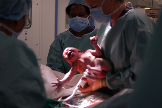
Approximately one-third of all babies born in the United States are currently delivered by cesarean section, according to the Centers for Disease Control and Prevention. Although cesarean delivery can be life saving for both the mother and child, the rapid increase in the cesarean birth rate between 1996 and 2011 raised significant concern that cesarean delivery is being overused.
This concern has led to initiatives to lower the c-section rates, including a new plan funded by the Oakland-based California HealthCare Foundation (CHCF) to lower California’s c-section rate for low-risk mothers to 23.9% in the next five years — in alignment with the Healthy People 2020’s national target.
A recent KQED Science article describes these efforts to reduce the state’s c-section rates. The story also explores the controversial issue that a healthy pregnant woman’s likelihood of having a cesarean birth varies depending on the hospital, based on a recent analysis of maternity care. For instance, the assessment report found that Lucille Packard Children’s Hospital Stanford has a c-section rate of 23.0 percent and the Coastal Communities Hospital in Santa Ana has a rate of 42.9 percent.
Deirdre Lyell, MD, professor of obstetrics and gynecology, clarified the issue in a recent email:
Nationally and internationally, there is concern that cesarean rates as a whole are too high. CHCF and others have shown a wide rage in cesarean rates by hospital around the country, and even within hospitals among individual physicians. Hospitals with very high rates should examine the underlying reasons. However, the “ideal rate” depends on the characteristics of the patient population, and it would be inappropriate to apply one goal to all women. For example, a pregnant, non-obese 25-year old who has had a prior vaginal delivery has a better likelihood of delivering her baby vaginally than does a pregnant, obese 45-year old first time mom.
At Stanford, we follow the “Safe Prevention of the Primary Cesarean Delivery” guidelines outlined by the American College of Obstetricians and Gynecologists and the Society for Maternal-Fetal Medicine. We care for a higher-risk maternal and higher-risk fetal population, and share with our patients a common goal for delivery: a safe mom and a safe baby, while not performing cesareans unnecessarily. Avoidance of the first cesarean helps reduce the potential risks in the future.
This is a reposting of my Scope blog story, courtesy of Stanford School of Medicine.