If you suffer from a very rare disease, getting the proper diagnosis can be an arduous journey. But a bigger challenge may be the feeling of isolation, since there may not be any support groups where you can connect to someone who is going through the same thing.
That was the situation the Bigelow family found themselves, and they turned to social media for the solution.
Bo Bigelow knew that his six-year-old daughter Tess had a genetic mutation called USP7. She also had global developmental delays in basic functions such as walking and talking, causing her to function at the level of an 18-month year old. Was USP7 the cause of her developmental delays?
Bigelow spread the word about his daughter’s genetic condition to find out, posting on Facebook, Twitter and a personal website with the plea to “help us find others like Tess.” A friend of the family also posted on Reddit, where it was read within 24 hours by a researcher at Baylor College of Medicine who was studying USP7. His research group had already identified seven children similarly affected by the same genetic mutation, and they were about to publish an article about it in Molecular Cell.
Tess may become part of future clinical trials at Baylor, but the researchers also connected the Bigelows to the other seven families. “These days there are ribbons and awareness-weeks for so many diseases,” Bigelow said in a recent KQED Science story, “but when yours is ultra-rare, you feel completed isolated. You feel like you’re never going to hear another person say, ‘Us too!’ And being connected to other families changes all that.”
The KQED piece goes on to explain:
“Patients or parents like Tess’ who are seeking answers to seemingly unsolvable medical mysteries have new tools to reach out, not only on social media, but in crowdsourcing websites like CrowdMed, a subscription service for people seeking answers to medical conundrums. At CrowdMed, people who have symptoms but have yet to find a diagnosis seek opinions from the site’s “medical detectives,” only some of whom are medical professionals.”
This is a reposting of my Scope blog story, courtesy of Stanford School of Medicine.
The hit new crime thriller Blindspotis about a mysterious woman, Jane Doe, who is covered in extensive full-body tattoos. If Jane Doe were a real woman who ever needed medical imaging, she might need to be concerned.
In a case report published recently in the journal Obstetrics & Gynecology, researchers found that extensive tattoos can mimic metastases on images from positron emission tomography (PET) fused with computed tomography (CT). PET-CT imaging is commonly used to detect cancer, determine whether the cancer has spread and guide treatment decisions. A false-positive finding can result in unnecessary or incorrect treatment.
Ramez N. Eskander, MD, assistant professor of obstetrics and gynecology at UC Irvine, and his colleagues describe the case study of a 32-year-old woman with cervical cancer and extensive tattoos. The pre-operative PET-CT scan using fluorine-18-deoxyglucose confirmed that there was a large cervical cancer mass, but the scan also identified two ileac lymph nodes as suspicious for metastatic disease. However, final pathology showed extensive deposition of tattoo ink and no malignant cells in those ileac lymph nodes.
It is believed that carbon particles in the tattoo pigment migrate to the nearby lymph nodes through macrophages, using mechanisms similar to those seen in malignant melanoma. The researchers explain in their case report:
Our literature search yielded case reports describing the migration of tattoo ink to regional lymph nodes in patients with breast cancer, melanoma, testicular seminoma, and vulvar squamous cell carcinoma, making it difficult to differentiate grossly between the pigment and the metastatic disease, resulting in unnecessary treatment.
The authors warn other physicians to be aware of the possible effects of tattoo ink on PET-CT findings when formulating treatment plans, particularly for patients with extensive tattoos.
This is a reposting of my Scope blog story, courtesy of Stanford School of Medicine.
The device on the golden fingertip is the skin-like sensor developed by Stanford engineers. (Bao Lab)
A hand without a sense of touch doesn’t really feel like a hand, many amputees describe. It’s more like a pliers that can be manipulated by sending signals from the brain to the prosthetic device. They dream of being able to delicately pick up a glass or to feel the touch of a loved one’s hand.
Stanford chemical engineering professor Zhenan Bao, PhD, and her team have spent a decade trying to help make this dream a reality, by developing a material that mimics skin and its sensory functions. Taking a big step towards this goal, they have now created a skin-like material that can tell the difference between a soft touch and a firm handshake.
Their artificial skin has two layers. The bottom layer acts as a circuit that transports pulses of electricity to nerve cells and translates these signals into biochemical stimuli that the nerve cells can detect. The top layer is a sensing mechanism composed of thin plastic embedded with billions of carbon nanotubes. When pressure is put on the plastic, the nanotubes are squeezed closer together enabling them to conduct electricity. What’s new is that the top layer can now detect pressure over the same range as human skin.
This allowed the plastic sensor to mimic human skin, which transmits pressure information to the brain as short pulses of electricity, similar to Morse code. Increasing pressure on the waffled nanotubes squeezes them even closer together, allowing more electricity to flow through the sensor, and those varied impulses are sent as short pulses to the sensing mechanism. Remove pressure, and the flow of pulses relaxes, indicating light touch. Remove all pressure and the pulses cease entirely.
A paper describing Bao’s new research has just been published in Science. As Bao comments in the release, “We have a lot of work to take this from experimental to practical applications. But after spending many years in this work, I now see a clear path where we can take our artificial skin.”
This is a repost of my Scope blog story, courtesy of Stanford School of Medicine.
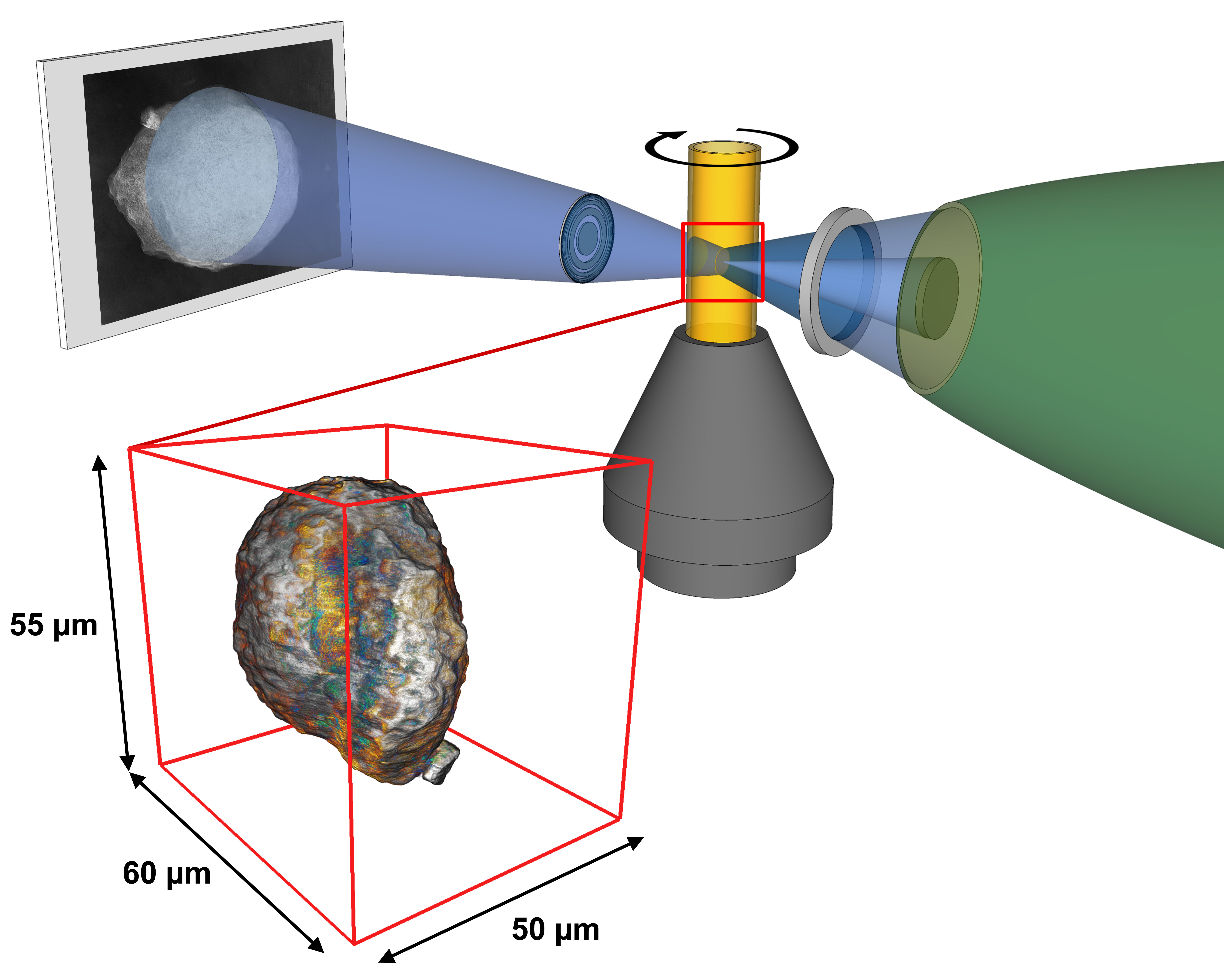
SSRL X-rays are focused to illuminate a small sample of catalysts inside a movable cylindrical holder. A lens magnifies the resulting sample image onto a screen, a CCD camera captures the 2-D image, and software is used to reconstruct a 3-D image of the single catalyst particle from a series of these 2-D images. (Florian Meirer/Utrecht University)
Scientists at SLAC and Utrecht University have identified key problems in the crude oil refining process in an effort to increase the production yield of gasoline.
Their recent experiments at SLAC National Accelerator Laboratory studied catalysts that crack apart the long-chain hydrocarbons in crude oil into smaller, more valuable hydrocarbons like gasoline. The efficiency of this refinement process decreases as the catalysts age.
The researchers used X-ray beams at SLAC’s Stanford Synchrotron Radiation Lightsource to image whole catalyst particles and their internal structure with high resolution – like taking a landscape photograph where you can see a panoramic view and zoom in to see the ants.
To learn more about this research, check out my communications article for SLAC National Accelerator Laboratory.
Morphine is a common narcotic pain reliever with significant side effects (sfxeric/Flickr).
Morphine is a powerful narcotic, commonly used to treat moderate to severe pain from surgery, injury and chronic health conditions like cancer or osteoarthritis. However, morphine has many negative side effects. It can cause drowsiness, nausea, vomiting, dizziness and constipation. More troubling, people can become dependent on it.
According to the Centers for Disease Control and Prevention, narcotic dependence and overdose deaths are a growing health problem in the United States. Narcotic sales quadrupled from 1999 to 2010 and narcotic-related deaths more than tripled from 1999 to 2012. More than 2 million people in the U.S. currently abuse narcotics.
In order to address this problem, researchers have long searched for alternative drugs that effectively relieve pain without inducing dependence as morphine does. A team of researchers now believe they’ve found a good candidate for such a drug, although it will be a long time before this drug, if it proves to be effective, is available in stores.
The LCLS creates the brightest X-ray source on the planet, which researchers need to accurately study the structure of tiny, nanoscale drug compounds.
How Narcotics Work
Narcotics are also called opioid pain relievers, because they block the sensation of pain by binding to opioid receptors within the pain pathway of the brain. A drug can provide pain relief by binding to one of three types of opioid receptors in the brain: delta, mu and kappa. Narcotics like morphine target the opioid mu receptors.
The problem is that long-term use of morphine reduces the number of available opioid mu receptors. As a result, tolerance develops so a higher dose of the drug is needed to achieve the same level of pain relief. Ultimately, prolonged use leads to physical dependence — which is when the neurons adapt to the presence of the drug and function normally only when it’s in the body.
Previous research has shown that administering morphine with another drug that simultaneously blocks the opioid delta receptors prevents this morphine-induced tolerance and dependence. Researchers just need to find the right drug combination with minimal side effects.
Recently, a team of researchers, led by University of Southern California chemistry professor Vadim Cherezov, developed a drug that activates opioid mu receptors while blocking opioid delta receptors. Their drug is derived from a specialized peptide – a naturally occurring chain of amino acids. Their research results were reported in the February issue of Nature Structural and Molecular Biology.
Studying Tiny Crystals
Unfortunately, it is very difficult to study new opioid compounds like the one Cherezov recently developed.
In order to understand the structure of new compounds, scientists usually grow crystals of the compound and then hit them with X-rays. By measuring the angles and intensity of how the X-rays bounce off the crystals, they can produce a three-dimensional picture of the crystal structure.
However, it can be difficult to grow crystals large enough to use this standard method, called X-ray crystallography. Plus, you typically need to freeze the crystal to make it more rigid, rather than study it in natural conditions and temperatures. On top of that, a conventional X-ray beam might blast and destroy the small crystal before you can collect enough data.
Instead, Cherezov’s team used the Linac Coherent Light Source to perform their experiment on tiny crystals at room temperature. LCLS X-ray pulses last just a quadrillionth of a second, or 100 times faster than it takes light to travel the width of a human hair. Yet they are a billion times brighter than a conventional X-ray source.
Using this unique X-ray source with higher-intensity, very short pulses, Cherezov was able to use smaller crystals and still collect terabytes of structural data before the crystals vaporized.
Each crystal the research team prepared was a millionth of a meter and contained many copies of their new opioid compound bound to an opioid receptor. The team placed the crystals in a toothpaste-like gel to simulate the receptors’ natural environment, and then injected a thin stream of this gel into the path of the LCLS X-ray beam.
The resulting structural model was precise enough to show how the new drug molecules bind with the receptor. This atomic-scale map should help scientists develop pain-relieving drugs with fewer negative side effects. It’s likely to to take years, though, before it can be tested in humans.
“This work will provide a solid basis for the design of a new generation of pain relievers with reduced dependency,” Cherezov said in a press release.
Cherezov’s experiment is just one of many performed at the Linac Coherent Light Source.
The LCLS is a Department of Energy User Facility where approximately 600 scientists conduct groundbreaking experiments each year, across many fields, including chemistry, biology, material science, technology and energy science.
LBNL Institute for Globally Transformative Technologies research team with prototype vaccine fridge and backpack for developing countries. (Berkeley Lab / Roy Kaltschmidt)
Vaccines are arguably one of the most important inventions of mankind. Unfortunately, vaccines must be produced and stored in an environment with very tight temperature regulation – between 36 °F and 46 °F – to keep the vaccine bugs alive. So vaccine delivery is a major problem due to the absence of reliable refrigeration in many remote countries.
Approximately 30 million children worldwide – roughly one in five – do not receive immunizations, leaving them at significant risk of disease. As a result, 1.5 million children under the age of five die annually from vaccine-preventable diseases, such as pneumonia and diarrhea. Perhaps more surprising, almost half of the vaccines in developing countries are thrown away because they get too warm during delivery so they are no longer viable. Some administered vaccines are also ineffective because they froze during transport, but there is no easy way to test this.
Scientists at Lawrence Berkeley National Laboratory (LBNL) are trying to solve this vaccine delivery problem by developing a portable solar-powered fridge. Fabricated entirely at LBNL, their portable solar-powered vaccine fridge will be transported by bicycle or motorcycle in remote areas of the developing world. Zach Friedman and Reshma Singh are leading the project as part of the LBNL Institute for Globally Transformative Technologies, which seeks to bring scientific and technological breakthroughs to address global poverty and related social ills.
The team’s first prototype portable fridge uses a thermoelectric heat pump, rather than a traditional vapor compression heat pump that relies on a circulating liquid refrigerant to absorb and remove heat. The thermoelectric chips were initially developed to keep laptops cool, so laptops could be made thinner without fans. The technology was adapted for this global application to reduce the size and weight of the fridge.
Their portable units have a one to three-liter capacity, much smaller than standard solar fridges that are typically 50 liters or more. Once the fridge cools down to the right temperature (36 °F – 46 °F), it is designed to run within that temperature range for at least five days without any power, at an ambient outside temperature as hot as 110 °F.
Before the researchers can field test their first prototype fridge in Africa, they need to pass the World Health Organization’s Performance, Quality and Safety testing protocol for products used in immunization programs. They are currently busy performing in-house testing at LBNL to ensure that they pass the formal tests, which will be conducted by an independent laboratory in the UK.
“We aren’t in the process of field testing yet, but we have established field testing agreements in both Kenya and Nigeria and have locations identified,” said Friedman. “We expect to start testing this coming year.”
Meanwhile, they are continuing their portable fridge development. “Currently, we are pursuing both thermoelectric and vapor compression heat pumps, even for these smaller devices,” explained Jonathan Slack, lead engineer. “It is not clear which will win out in terms of manufacturability and affordability.”
They are also developing a backpack version of the vaccine fridge. However, human-carried devices have to meet stricter World Health Organization standards, so they are focusing at this stage on the small portable fridge instead.
Ultimately their goal is to make it easy for health care workers to deliver viable vaccines to children in remote areas, solving the “last miles” of vaccine delivery.
Eyeglasses may no longer be necessary to see computer screens. (F H Mira, flickr)
What if everyone could clearly see their smart phone, tablet, computer and TV screens without having to wear corrective eyeglasses or contact lenses?
Approximately 75% of American adults use some form of corrective lenses to see or read properly. And most of us need them to see computer screens on a daily basis. Now researchers are developing new technology that uses computer algorithms to compensate for an individual’s visual impairment, so many of us may soon be able to ditch our glasses and contacts.
Brian Barsky, UC Berkeley professor of computer science and vision science and affiliate professor of optometry, teamed up with colleagues at UC Berkeley and MIT to improve vision-correcting display technology. They developed a combination of hardware and software improvements to achieve both high image resolution and contrast simultaneously, a major milestone. Their results were recently published in a paper in the ACM Transactions on Graphics.
First, they modified an iPod touchscreen by adding a standard light field display – a mask with an array of pinholes sandwiched between thin layers of plastic. The tiny pinholes were each 75 micrometers in diameter and spaced 390 micrometers apart. This light field display was used to enhance image contrast, providing a full range of bright colors in the displayed images.
The researchers also developed complex, innovative computer algorithms to adjust the light intensity from each pinhole. These algorithms helped enhance the resolution or sharpness of the displayed images. The researchers can use a person’s eyeglass prescription to compute an altered image, that when viewed through the light field display, appears in sharp focus for that individual.
“Our technique distorts the image such that, when the intended user looks at the screen, the image will appear sharp to that particular viewer,” said Barsky in a press release. “But if someone else were to look at the image, it would look bad.”
The technology could not only help the millions of people who wear glasses and contacts, but also those with complex vision problems that cannot be corrected. The most common vision problems – nearsightedness, farsightedness and astigmatism – are usually easily corrected with standard lenses. However, people with complex vision problems often have irregularities in the shape of their eyes’ surface or cornea, requiring new kinds of corrective lenses that are still under development.
“We now live in a world where displays are ubiquitous, and being able to interact with displays is taken for granted,” said Barsky. “People who are unable to view displays are at a disadvantage in the workplace and life in general, so this research could transform their lives.”
In the future, the researchers plan to incorporate commercially available eye trackers to adapt the displayed images to the user’s head position. They also hope to develop display screens that appear clear to multiple users with different visual problems.
Scientists are developing a portable device that can measure a person’s radiation exposure in minutes. This image shows a magneto-nanosensor chip reader station, chip cartridge, and chip. (Credit: S. Wang)
Picture the scene of the Fukushima nuclear accident. The Daiichi nuclear reactors were hit by an earthquake of magnitude 9.0 and flooded by the resulting tsunami, which caused a nuclear meltdown and release of radioactive materials. Over 100,000 people were evacuated from their homes due to the threat of radiation contamination.
In a large-scale radiological incident like this, emergency medical personnel need a rapid way to assess radiation exposure so they can identify the people who need immediate care. This radiation-dosimetry technology needs to be sensitive, accurate, fast and easy to use in a non-clinical setting.
Local scientists have developed a small, portable device that can quickly test the level of radiation exposure victims have suffered in such emergencies. This technology was developed by scientists from Berkeley Lab, Stanford University and several other institutions, as reported in a journal article recently published in Scientific Reports. The lead researchers were Dr. Shan Wang from Stanford University and Dr. Andrew Wyrobek from Berkeley Lab.
This new dosimetry device is a novel type of immunoassay. Immunoassays are chemical tests used to detect or measure the quantity of a specific substance in a body fluid sample using a reaction of the immune system. For example, a common immunoassay test for pregnancy measures the concentration of the human chorionic gonadotropin hormone in a woman’s blood or urine sample.
In order to measure a person’s radiation dose, the new device measures a blood sample for the concentration of particular proteins that change after radiation exposure. Scientists, including those in Wyrobek’s group, have previously identified these target proteins as excellent biological markers for radiation dosimetry. Basically, blood exposed to radiation has a special biochemical signature.
But scientists needed more than just target proteins. They also needed an accurate, sensitive way to quickly measure the proteins’ concentrations in a few drops of blood. So at the heart of the new device is a biochip developed by Wang’s group.
The biochip system relies on a sandwich structure where a target protein is trapped between a capture antibody and a detection antibody. The capture antibodies are immobilized on the surface of the biochip sensor. When a drop of blood is placed on the biochip, those antibodies capture the target proteins and the other proteins are washed away. Detection antibodies labeled with magnetic nanoparticles are then added, forming a sandwich structure that traps the target proteins. When an external oscillating magnetic field is then applied, the magnetic nanoparticles generate an electrical signal that is read out. This signal measures the number of magnetic nanoparticles bound to the surface, and this indicates the number of target proteins that have been trapped.
The researchers tested the biochip system using blood from mice that had been exposed to varying levels of radiation. Their novel immunoassay results were validated by comparing them to conventional ELISA immunoassay measurements. Overall the scientists demonstrated that the new biochip dosimetry system is fast, accurate, sensitive and robust. In addition, the whole system is the size of a shoebox so it is very portable.
“You add a drop of blood, wait a few minutes, and get results,” explained Wyrobek in a press release. “The chip could lead to a much-needed way to quickly triage people after possible radiation exposure.” Although the technology is still under development, hopefully it will be available before the next radiological accident or terrorist attack occurs.
For more information about this biochip system, check out my KQED Science blog.
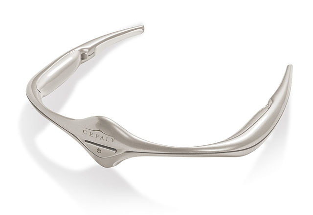
Photograph of Cefaly anti-migraine device, courtesy of STX-Med via Creative Commons license
While shopping for groceries at Trader Joes, suddenly your peripheral vision disappears. This could be frightening, but you know what is coming — a one-sided pulsating pain, sensitivity to light and noise, nausea, vomiting and seeing flashing lights. You quickly drive home and cancel your plans, because you have a migraine coming. You need to lie still in a dark quiet room for the next 24 hours.
Migraines affect about 30 million Americans. This means that one in four households in the US have at least one member impaired by migraines. Women are three times more likely to be migraine sufferers than men.
Unfortunately, there is currently no cure for migraines. A migraine diary can help identify the headache triggers to avoid. Medications can also help reduce the number of attacks or ease the symptoms, but these medications are often ineffective or cause unpleasant side effects.
Instead migraine sufferers might find relief from a new non-medicinal alternative, a device called a supraorbital transcutaneous stimulator (STS) that stimulates the nerves around the eyes and forehead. A study recently published in Neurology tested the safety and effectiveness of this STS device designed to prevent migraines.
Conducted by researchers in five specialized headache clinics in Belgium, this study was a randomized controlled trial that compared the STS device with an identical-looking sham device. Study participants were aged 18 to 65 who routinely experienced a minimum of two migraine attacks per month. None of the 67 participants had taken anti-migraine medications in the three months leading up to the study.
Both the STS and sham devices used a self-adhesive electrode placed on the forehead that buzzed identically during treatment. Only the STS devices delivered electrical impulses. The participants wore one of the devices for 20 minutes per day for 90 days.
The participants’ migraine diaries indicated that the number of migraine attacks dropped by at least half for 38% of the participants using the STS device, compared with 12% for those using the sham device. Although the severity of the migraines was not reduced, people using the STS device had fewer days with headache, fewer total migraine attacks, and used fewer pain relief medications each month. Most importantly, there were no adverse effects seen in either group.
The study concluded that treatment with a STS device is “effective and safe as a preventive therapy for migraine.” However, only 67 migraine sufferers have been studied and the use of this device was only examined for three months. Larger studies with longer-term treatment are needed to confirm that this STS device is safe and effective.
For more information about migraines and the STS device, check out my KQED Quest blog.
Artist’s animation depicting the moment that NASA’s Curiosity rover touches down onto Mars. (NASA/JPL-Caltech image)
When I tried to make lunch plans with a friend for next week, he didn’t know yet whether he could meet me. That’s because his plans depend on how smoothly the Curiosity rover lands on Mars tonight. His research team put together the Radiation Assessment Detector that is mounted on the top deck of the Curiosity rover.
NASA’s Mars Science Laboratory spacecraft with the Curiosity rover are approaching Mars at this moment. It’s expected to land tonight at 10:31 p.m. PDT (Pacific Daylight Time). The technical challenges involved in the Curiosity’s landing are daunting. The final minutes to landing are described beautifully in the NASA Jet Propulsion Laboratory’s popular video dubbed “The Seven Minutes of Terror.”
We still aren’t sure if life ever existed on Mars. From past missions, researchers know that there used to be water there. Now they want to determine if Mars once had the kind of environment that could be habitable or conducive for the formation of microbial life.
The Curiosity rover is a car-like rover that will search Mars for past or present conditions favorable for life on the planet. It is basically a science lab on wheels, including 10 complex scientific instruments. These instruments are designed to study the chemistry of rocks, soil and atmosphere — searching for signs of past life on Mars.
One of those scientific instruments is the Radiation Assessment Detector, which is designed to characterize the energetic particle spectrum at the surface of Mars. This will allow researchers to determine the radiation dose for humans at the planet surface, as well as provide input on the effects of particles on the surface and atmosphere. The surface is thought to have much higher radiation than Earth, since Mars has a thinner atmosphere and no global magnetic shield to divert charged particles.
Although all research requires patience, hurling your research instrument at a far away planet requires both patience and guts. The landing may cause 7 minutes of terror, but the days of waiting must include its own nail-biting nervousness. When I get together with my friend for lunch, I’ll check his nails. Hopefully the landing will be a success, so he’ll be at the Jet Propulsion Laboratory for the next couple weeks though. I can wait.