Surgery to find your voice: A Q&A with expert Anna Messner

5540462170_5ca4645115_o_flickr_HowardLake_560x.315jpg
Photo by Howard Lake

When we’re in a noisy restaurant, it’s really difficult to hear my young niece speak. She can only talk very quietly, because she has a paralyzed vocal cord.

Like many children born very premature, the nerve going to her vocal cord was likely damaged when she had heart surgery soon after she was born. Her inability to be heard frustrates her, especially now that she is in school. However, a rare surgery may bring her the hope of a near-normal voice.

Stanford surgeons recently began performing laryngeal reinnervation surgery, which essentially rewires the paralyzed vocal cord with a new nerve supply. I recently spoke with Anna Messner, MD, a professor of otolaryngology and pediatrics who sees patients at Lucile Packard Children’s Hospital Stanford, about laryngeal reinnervation surgery.

What standard surgical procedures are used to treat unilateral vocal cord paralysis?

In general, the surgical procedures bulk up the paralyzed vocal cord to move it towards the midline of the body, making it easier for the other vocal cord to compensate and close. There are two standard surgeries. We can do injection laryngoplasty, where we inject a substance into the paralyzed vocal cord to thicken it. Unfortunately, this procedure often needs to be repeated multiple times, if it works at all. We can also insert a medialization implant in teenagers and adults, but this doesn’t work for growing kids. If we put an implant into a two year old, it wouldn’t be an appropriate size when he is 10.

How does laryngeal reinnervation surgery work?

No matter what we do, we can’t make the vocal cord move. We can never make it perfect again. What we can do is hook up one of the other nerves in the neck to the recurrent laryngeal nerve that goes to the vocal cord. And that helps some new nerve fibers go to the vocal cord, making the vocal cord stronger and thicker. As a result, the voices on these kids improve significantly.

The surgery itself is fairly straightforward and only takes about an hour. The children typical go home the same day or just stay overnight, and they feel back to normal in a couple of days. But then we have to wait five to six months for the nerve fibers to grow before we can see real improvement in the voice. The only downside is that it takes a long time to see the effects of the surgery.

What inspired you to learn the laryngeal reinnervation procedure?

We have a large pediatric cardiac surgery program at Stanford, so we have quite a few patients with vocal cord paralysis. Most of our patients are born prematurely and need heart surgery, which can pull and damage the nerve that goes to the vocal cord on one side. After these surgeries, the damaged vocal cord starts working again in just over a third of the cases. But for the rest of the kids, the vocal cord remains paralyzed.

The standard surgeries just don’t work very well, so we’ve had a longstanding interest in finding alternatives. I saw Marshall Smith, MD, the medical director of the Voice Disorders Center at University of Utah, give a presentation on his clinical trials. So I observed him performing the reinnervation surgery about 1.5 years ago, and since then I’ve been performing the surgery. One of my colleagues, Doug Sidell, MD, also performs the surgery.

How does the voice improvement impact the patients?

The voice improvement makes a huge impact on the children, especially in school. For instance, when the children are trying to read a story or give a presentation in front of the classroom, now they can actually be heard. The results are very encouraging. The surgery has the potential for huge, life-long voice improvement.

This is a reposting of my Scope medical blog story, courtesy of Stanford School of Medicine.

Should doctors give up their white coats?

Photo by NEC Corporation of America

When you google ‘doctor,’ virtually every image shows a person in a long-sleeved white coat. The crisp white coat with a stethoscope around the neck has long symbolized the profession. However, there is currently controversy about whether doctors should give up their classic uniform, as described in a recent story in the Boston Globe.

Britain’s National Health Service banned white coats back in 2008, requiring doctors to be bare below the elbows to avoid spreading infections. Many clinical departments in the United States have done the same. The argument goes something like this:

  • The sleeves of white coats are germ magnets.
  • Doctor’s don’t launder their white coats very often, so deadly infections can be spread from one patient to another.
  • Therefore, doctors shouldn’t wear long-sleeved white coats.

As a pediatric infectious disease specialist at Stanford, Charles Prober, MD, supports this theory:

In terms of the infectious disease risk, there is little question that one can carry bacteria or viruses on your clothing — whether it’s a white coat or the sleeve of your shirt. And one way to lessen that is to wash up to the elbows, especially when you’re going into high-risk environments like the ICU or nursery. Obviously you can only wash up to your elbows if they aren’t covered with something.

William Benitz, MD, division chief of neonatal and developmental medicine, agrees:

I find the summary reports highly credible and accept the contention that the long sleeves of white coats harbor infectious agents and carry them from patient to patient. We banned white coats in our NICU about 5 years ago, along with a mandate for baring arms to the elbow and hand cleansing upon entering any patient room. Part of the reason for the former is to reinforce and provide active visual evidence of the latter. We used to hear ‘but I won’t touch anything’ a lot, but that was often not so. Not an issue now.

However, there are some practical reasons for wearing white coats and not even Prober has given his up. He explains:

When I go to the hospital, I wear my white coat over there because it allows me to carry a bunch of stuff in my pockets. Otherwise, I’d have to carry it in my hands. But I usually take the coat off when I’m seeing patients. It’s said that some children are frightened when they see the white coat. I normally just wear a long-sleeved shirt, tie and pants so I roll my sleeves up to my elbows when I go into the nursery. I wear bow ties and argue that they are less likely to have bacteria than straight ties, plus a child can’t easily grab a bow tie and soil it.

Many doctors aren’t ready to give up there white coats though, so the debate is likely to continue. Prober estimates, “Walking up and down the hall, the number of people wearing white coats at Stanford hospital is probably about 50 percent.”

This is a reposting of my Scope blog story, courtesy of Stanford School of Medicine.

A tiny fish helps solve how genes influence longevity

Photograph of the Nothobranchiius furzeri killifish, by Ugua

A tiny, short-lived fish may help solve one of the largest mysteries: how do genes influence longevity?

The African turquoise killifish has evolved as the shortest-lived known vertebrate — driven by its survival in the hot climate of Mozambique and Zimbabwe in seasonal ponds that only exist for a few months during the wet season. This compressed life span makes the killifish an ideal organism for genetic studies on aging and longevity.

To help researchers study this intriguing animal model, Stanford geneticist Anne Brunet, PhD, and her colleagues have now fully mapped the genome of the African turquoise killifish. Their initial insights into the genetic determinants of the killifish’s life span were published today in Cell.

Brunet’s team sequenced short segments of the killifish DNA and then assembled them using specialized software to create a complete map of the turquoise killifish genome.

Brunet explains in a news release:

The range of life spans seen in nature is truly astonishing, and really we have very little insight into how this has evolved or how this works. By having the genome of this fish and comparing it to other species, we start seeing differences that could underlie life span differences both between species and also within a species.

In the article, the researchers report on their initial study of genes unique to the short-lived killifish, which were identified by comparison to longer-lived species, such as killifish that were mated with longer-lived fish. Surprisingly, they found that the genes associated with life span differences between various killifish strains are clustered on the sex chromosomes, so its short life span likely co-evolved with sex determination. They also identified some unusual aging genes in both killifish and other long-lived fish, raising the question of what role these aging genes play in the determination of life span.

To uncover all of the killifish’s traits, Brunet’s group will have help: They have created a user-friendly website, which provides other researchers with free access to the data.

Brunet explained in the news release, “They can go to our website, enter their favorite gene of interest, and then zoom in on the killifish equivalent.”

This is a reposting of my Scope blog story, courtesy of Stanford School of Medicine.

Walking tall: the challenge of correcting your gait

Photo by sean_hickin

Thanks to a bum knee, in elementary school I had to choose between two styles of special oxford shoes to help correct my gait; I hated those ugly shoes. Luckily these days I have many cute options, along with custom orthotics.

Despite good shoes though, my plantar fasciitis recently sent me back to my physical therapist. Of course the first thing she had me do is walk back and forth across the room, a common sight at any physical therapy office.

Now everyone from doctors and physical therapists to yoga instructors are teaching people how to walk properly.

A recent story in Vogue chronicles one woman’s efforts to correct her bad habits at two walking and gait clinics — with a physical therapist in Santa Monica and with a yoga teacher in Brooklyn. Author Marisa Meltzer also checked in with Jessica Rose, MD, a professor of orthopedic surgery at Stanford University and director of the Motion & Gait Analysis Laboratory at Lucile Packard Children’s Hospital.

Meltzer received immediate feedback from the physical therapist, displeased with the uneven wear of her shoes. As Meltzer describes:

Finally, she instructs me to walk back and forth across the room. Her diagnosis: I’m constantly leaning back like a Looney Tunes character approaching oncoming traffic.

Similarly, Meltzer visits well-known yoga teacher, learning to straighten her upper body, rotate her pelvis, and swing her arms as she walks. She describes her new gait near the end of the article: “It feels unnatural, yet when I catch my reflection in the mirror I see I’m moving elegantly and with confidence.”

This is a reposting of my Scope blog story, courtesy of Stanford School of Medicine.

A cure is not enough for young cancer survivors

Photo by klimkin
Photo by klimkin

I survived Hodgkin’s lymphoma as a young adult about twenty years ago, thanks to the chemotherapy and radiotherapy that I received at Stanford Hospital as part of a clinical trial.

Even back then, the focus of the research was on fine-tuning my cancer treatment to maintain an excellent likelihood of survival, while minimizing the long-term health problems due to therapy. I knew Hodgkin’s was unlikely to kill me, so I had to worry instead about future health issues caused by my radiation and chemotherapy.

People that survive cancer at a young age are expected to live many decades after diagnosis and treatment, so they are the most vulnerable population to long-term damaging effects from cancer therapy. Stanford’s Karen Effinger, MD, MS, and Michael Link, MD, explore this issue in an editorial published today in JAMA Oncology.

The editorial explains that it is critical to directly study the late effects in young adult cancer survivors, rather than the common practice of extrapolating from studies of children and middle-aged adults.

In particular, they discuss a new study by Katherine Rugbjerg, PhD, and Jorgen Olsen, MD, DMSc, from the Danish Cancer Society Research Center, which used the national Danish registries to compare the long-term risk of hospitalization in almost 34,000 5-year survivors of adolescent and young adult cancers with that of more than 228,000 age- and sex-matched population controls. Reported in the latest issue of JAMA Oncology, Rugbjerg and Olsen found that adolescent and young adult cancer survivors had significant health issues due to their treatment; however, these treatment effects were different than survivors of childhood cancers.

The editorial also discusses the late effects of pediatric cancer treatment on survivors’ neurocognitive development, which impacts education, employment and quality of life. Effinger and Link specifically describe a new study reported in JAMA Oncology by Kevin Krull, PhD, and colleagues from the St. Jude Children’s Research Hospital, which compared the neurocognitive outcomes in 80 adult 25-year survivors of a pediatric cancer with 39 controls. Krull concluded that the risk of neurocognitive impairment from cancer treatment was related to the development of chronic health conditions — rather than directly from exposure to high-doses of chemotherapy, as expected — but longitudinal studies are needed to sort out possible modifying factors.

The editorial authors conclude:

Going forward, we must apply our knowledge of late effects to improve monitoring and interventions for patients. While the progress made in the management of cancer in children and young adults has been gratifying, we must remember the words of Giulio D’Angio, who reminds us that “cure is not enough.”

This is a reposting of my Scope blog story, courtesy of Stanford School of Medicine.

Researchers develop molecular target for brain cancer

Images by Weibo Cai/Department of Radiology, University of Wisconsin-Madison. On the left, the antibody is linked to a label that shows up in a PET scanner, and the aggressive cancer shines brightly. On the right, a similar cancer without the molecular marker is less obvious.
Images by Weibo Cai/Department of Radiology, University of Wisconsin-Madison. On the left, the antibody is linked to a label that shows up in a PET scanner, and the aggressive cancer shines brightly. On the right, a similar cancer without the molecular marker is less obvious in the PET scan.

About 23,000 new cases of brain and central nervous system tumors are diagnosed annually, and more than 15,000 patients are expected to die of brain cancer this year in the United States, according to the American Cancer Society. Glioblastoma multiforme is the most common brain malignancy, but it remains incurable with only 5% of patients surviving at least 5 years after diagnosis. This bleak scenario has motivated the search for a better molecular target for glioblastoma multiforme diagnosis and therapy.

Weibo Cai, PhD, associate professor of radiology and medical physics, and his research team at the University of Wisconsin-Madison searched the Cancer Genome Atlas database and identified an effective biomarker for the deadly glioblastoma multiforme: the CD146 gene, which is highly active in glioblastoma.

CD146 genes place unique CD146 proteins on the surface of cells. Cai’s team developed an antibody that selectively latches onto the CD146 proteins concentrated on the glioblastoma tumors. They also tagged the antibody with a radioactive copper isotope, so the tumors could be easily identified and localized with a positron emission tomograph (PET), an imaging scanner commonly used to detect cancer.

Cai tested their antibody by implanting mice with human glioblastoma tumors, injecting them with the antibody and imaging them with a small animal PET scanner. The copper-labeled antibody preferentially accumulated in the tumors, allowing PET imaging to accurately identify tumors as small as 2 mm. Their study results were recently reported in the Proceedings of the National Academy of Sciences.

Cai explained in a university news release:

We’ve created a tag that – at least in our mouse model – is highly specific for this aggressive brain cancer. If the technique proves out in further tests, it could be used to diagnose some strains of aggressive glioblastoma, and also to evaluate treatment progress or even to test potential drugs.

The researchers also found high activity of CD146 in ovarian, liver, and lung tumors so their antibody could have a wide range of applications. However, there is a lot of research to be done before the technique could be used in the clinic. Cai said in the news release, “This targets tumors with the worst survival, but I want to emphasize that human trials are some years in the future.”

This is a reposting of my Scope blog story, courtesy of Stanford School of Medicine.

Stanford alumnus writes children’s book to inspire the next generation of curious minds

ALandrigan_Rose_B_cell_birthday_cake_560x324

Soon there will be a new superhero children’s book available, but these superheroes aren’t from Marvel comics. The book, Rose’s Superhero Birthday: An Immune Cell Treasure Hunt, is about the immune cell superheroes that keep us healthy.

Angela Landrigan, PhD, did her graduate and postdoctoral training in immunology at Stanford’s medical school, where she studied how immune cells respond to cancer. She now works at a private company that develops software used to analyze immunology “big data.” She’s also a busy mom to two energetic, curious girls, which led her to write and illustrate a new children’s book to make learning about the immune system fun. I spoke with Angela last week about her new book, which she plans to distribute on her website.

What inspired you to write a children’s book?

My kids led me to write this book, particularly my 4-year-old Violet. Sometimes I work from home analyzing datasets, and she’ll look over my shoulder and ask me all these deep questions about cells and what they do. Plus we talk through the details of everyday things, like if she gets a cut or flu shot. I realized that kids can pick up a startling amount of detail, and they’re so thirsty and eager for knowledge.

So I wrote the book to answer Violet’s questions, then I quickly realized that I have the opportunity to teach more children and even parents and caregivers about how our immune cells work. Immunology is becoming an increasingly popular topic in the public health conversation — anything from vaccine awareness to disease epidemics. My book can help people to have less fear of the unknown and to be better equipped to make decisions that influence their own lives and public health.

How did you develop the characters and storyline for your book?

 The main character emerged because my daughter Violet wanted me to tell her new stories every night before bed. So I created this character Rose who goes on adventures.

The book follows a 7-year-old girl named Rose, who is really excited about science. She asks her immunologist-Mom for a science-themed birthday party with a B-cell birthday cake and a treasure hunt for stuffed animal immune cells. The next day, Rose invites all her friends over for a play date to create and act out a play on how immune cells work together in concert to get rid of a virus.

I’ve tried to capture the joy of creation, exploration and discovery of childhood, while engaging kids to thinker deeper.

Who is your target audience?

My target audience is families with children – mostly children between 4-10 years old, but along the way adults will learn too. And it’s often parents who make the decisions that influence public health.

I’ve tried to include characters with diverse demographics — boys and girls of multiple races. Hopefully readers can identify with the characters no matter who they are.

ALandrigan_B_cell_card_200x280

Why do you think your book is important and what do your daughters think of it?

This book helps bridge the communication gap between all that we’ve learned as researchers and what’s being communicated to the public. Having been a research immunologist, I feel like there is a deficit in what’s getting communicated.

The book also inspires kids to ask questions and dig deeper. Perhaps these kids will go on to discover the next generation of cures or become creative problem solvers in other ways. I want to capitalize on this time when kids are sponges, excited and eager to learn, by planting the seeds of curiosity about science and the world.

I particularly want to empower girls in science. I hope to inspire girls to identify with Rose, so science doesn’t feel like a topic they have to shy away from – and show them that you can be a mom and still do science. The power of role models is tremendous.

It’s been a fun, inclusive family project — not an extra demand that required carving out time away from my kids. My daughters have been really excited along the way to read the drafts over and over.

Are there any more books in your future?

Yes, I’m thinking of writing another Rose book — a “take-your-daughter-to-work” book where Rose goes to the lab with her Mom to learn about how scientists are manipulating the immune system for therapeutic benefit or investigational purposes. I’m also planning to write and illustrate a baby board book on immune cells called “Bedtime for B-Cell.” Another book in the concept phase is an encyclopedia with less narrative, covering topics like allergies, vaccines and asthma. These other books will build upon the concepts of my first book, delving deeper into the details.

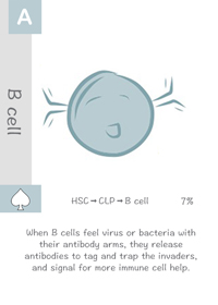
Illustrations from Rose’s Superhero Birthday: An Immune Cell Treasure Hunt by Angela Landrigan

This is a repost of my Scope blog story, courtesy of Stanford School of Medicine.

Stanford expert calls for more targeted anti-obesity policies

U. S. Department of Agriculture's helps educate shoppers about the value of food labeling in December 1975. Photo courtesy National Archives and Records Administration.
U. S. Department of Agriculture helps educate shoppers about the value of food labeling in 1975. Photo courtesy National Archives and Records Administration.

Reality TV shows like “The Biggest Loser” are popular in part because the audience can relate to the participants — more than two-thirds of adults and about one-third of children and adolescents are obese or overweight in the US. The Surgeon General and the Centers for Disease Control and Prevent have declared obesity to be a national epidemic and a major contributor to leading causes of death, including heart disease, stroke, diabetes and some types of cancer.

Although our country is committed to finding solutions to the increase in obesity, public policies have fallen short, according to Deborah Rhode, JD, a Stanford law professor and legal ethics scholar. In a recent journal article, she wrote:

Many policy responses have proven controversial, and those most often recommended have frequently faced an uphill battle at the federal, state, and local level. At the same time that obesity rates have been rising sharply, many jurisdictions have resisted, or rolled back, strategies such as soda taxes or regulation of advertising directed at children.

In the journal article, Rhode evaluates anti-obesity policies, including calorie disclosure requirements, taxes or bans on sugar-sweetened beverages, food stamp modifications, zoning regulations, children’s marketing restrictions, physical activities initiatives, food policies and education. She suggests that a more targeted approach is needed to combat obesity. For instance, Rhode recommends creative zoning regulations that restrict the location of fast-food restaurants near schools while encouraging healthy food retailers in underserved neighborhoods.

In a Stanford news release, Rhode noted that the first lady Michelle Obama’s “Let’s Move!” campaign against childhood obesity applies to politics as well as physical activity. Rhode summarized, “Although we need more evaluation of policy strategies, we know enough about what works to chart a course of reform. We should act now on what we know.”

This is a reposting of my Scope blog story, courtesy of Stanford School of Medicine.

Tattoo ink may mimic cancer on PET-CT images

19824000898_7611622d67_o_Flickr_PauloGuereta
Photograph by Paulo Guereta

The hit new crime thriller Blindspot is about a mysterious woman, Jane Doe, who is covered in extensive full-body tattoos. If Jane Doe were a real woman who ever needed medical imaging, she might need to be concerned.

In a case report published recently in the journal Obstetrics & Gynecology, researchers found that extensive tattoos can mimic metastases on images from positron emission tomography (PET) fused with computed tomography (CT). PET-CT imaging is commonly used to detect cancer, determine whether the cancer has spread and guide treatment decisions. A false-positive finding can result in unnecessary or incorrect treatment.

Ramez N. Eskander, MD, assistant professor of obstetrics and gynecology at UC Irvine, and his colleagues describe the case study of a 32-year-old woman with cervical cancer and extensive tattoos. The pre-operative PET-CT scan using fluorine-18-deoxyglucose confirmed that there was a large cervical cancer mass, but the scan also identified two ileac lymph nodes as suspicious for metastatic disease. However, final pathology showed extensive deposition of tattoo ink and no malignant cells in those ileac lymph nodes.

It is believed that carbon particles in the tattoo pigment migrate to the nearby lymph nodes through macrophages, using mechanisms similar to those seen in malignant melanoma. The researchers explain in their case report:

Our literature search yielded case reports describing the migration of tattoo ink to regional lymph nodes in patients with breast cancer, melanoma, testicular seminoma, and vulvar squamous cell carcinoma, making it difficult to differentiate grossly between the pigment and the metastatic disease, resulting in unnecessary treatment.

The authors warn other physicians to be aware of the possible effects of tattoo ink on PET-CT findings when formulating treatment plans, particularly for patients with extensive tattoos.

This is a reposting of my Scope blog story, courtesy of Stanford School of Medicine.

Is it time to compensate kidney donors?

Kidney transplant surgery (Tareq Salahuddin)

A recent New York Times blog entry editorialized on the worldwide shortage of transplant kidneys, raising the question of whether it’s time to compensate kidney donors to meet the growing need. The blog echoed the debate that is emerging in the United States among some doctors, medical societies, and groups that oversee organ transplants.

Taboos against paying for transplant organs are powerful. But these may be overcome by necessity, since the demand for transplant kidneys is growing at an alarming rate largely due to kidney failure from diabetes, high blood pressure and obesity-related diseases. According to the National Kidney Foundation, 450,000 Americans are on dialysis and the severe shortage of transplant kidneys in the U.S. results in 12 patient deaths each day.

Laying the groundwork for change, a collaboration of nephrology and finance experts, including Philip J. Held, PhD, a Stanford consulting professor of nephrology, performed a comprehensive cost-benefit analysis of a proposed government program for kidney donor compensation. In a study published last week in the American Journal of Transplantation, the authors estimate the shortage of transplant kidneys would be eliminated within five years if the government compensates living kidney donors $45,000 and the estates of deceased donors $10,000. The proposed compensation would also include an insurance policy against any health problems that might result from the donation.

The authors’ analysis shows that the benefits of a donor compensation program would greatly exceed the costs for society in general and taxpayers in particular. The researchers calculate the monetary value of a longer and healthier life for each kidney recipient at $1.3 million, with the added bonus of saving $1.5 million for not needing expensive dialysis treatments. After subtracting from these benefits the cost of transplants, society would enjoy a net welfare gain of $1.9 million over the lifetime of each kidney recipient. Since taxpayers currently pay about 75 percent of the cost of both dialysis and kidney transplants, this represents a taxpayer savings of about $400,000 per kidney recipient.

One of the main arguments against kidney donor compensation is that rich people would buy kidneys from poor people, exploiting them and causing them harm. The authors argue that the opposite is true because the poor, especially poor African Americans, are overrepresented on the kidney waiting list – so they would enjoy the greatest benefit.

The researchers summarized their findings in a press release supplied to our office:

“In sum, having the government compensate kidney donors would be a win-win-win situation. Kidney recipients would enjoy much longer and healthier lives. Kidney donors would receive compensation for their gift of life, whereas now they receive nothing. And taxpayers would save money because transplantation is not only a more effective treatment for kidney failure than dialysis, it is a much less expensive one.”

This is a repost of my Scope blog story, courtesy of Stanford School of Medicine.