Science news, education and opinions in plain English
Author: Jennifer Huber
As a Ph.D. physicist and research scientist at the Lawrence Berkeley National Laboratory, I gained extensive experience in medical imaging and technical writing. Now, I am a full-time freelance science writer, editor and science-writing instructor. I've lived in the San Francisco Bay Area most of my life and I frequently enjoy the eclectic cultural, culinary and outdoor activities available in the area.
U. S. Department of Agriculture helps educate shoppers about the value of food labeling in 1975. Photo courtesy National Archives and Records Administration.
Reality TV shows like “The Biggest Loser” are popular in part because the audience can relate to the participants — more than two-thirds of adults and about one-third of children and adolescents are obese or overweight in the US. The Surgeon General and the Centers for Disease Control and Prevent have declared obesity to be a national epidemic and a major contributor to leading causes of death, including heart disease, stroke, diabetes and some types of cancer.
Although our country is committed to finding solutions to the increase in obesity, public policies have fallen short, according to Deborah Rhode, JD, a Stanford law professor and legal ethics scholar. In a recent journal article, she wrote:
Many policy responses have proven controversial, and those most often recommended have frequently faced an uphill battle at the federal, state, and local level. At the same time that obesity rates have been rising sharply, many jurisdictions have resisted, or rolled back, strategies such as soda taxes or regulation of advertising directed at children.
In the journal article, Rhode evaluates anti-obesity policies, including calorie disclosure requirements, taxes or bans on sugar-sweetened beverages, food stamp modifications, zoning regulations, children’s marketing restrictions, physical activities initiatives, food policies and education. She suggests that a more targeted approach is needed to combat obesity. For instance, Rhode recommends creative zoning regulations that restrict the location of fast-food restaurants near schools while encouraging healthy food retailers in underserved neighborhoods.
In a Stanford news release, Rhode noted that the first lady Michelle Obama’s “Let’s Move!” campaign against childhood obesity applies to politics as well as physical activity. Rhode summarized, “Although we need more evaluation of policy strategies, we know enough about what works to chart a course of reform. We should act now on what we know.”
This is a reposting of my Scope blog story, courtesy of Stanford School of Medicine.
The hit new crime thriller Blindspotis about a mysterious woman, Jane Doe, who is covered in extensive full-body tattoos. If Jane Doe were a real woman who ever needed medical imaging, she might need to be concerned.
In a case report published recently in the journal Obstetrics & Gynecology, researchers found that extensive tattoos can mimic metastases on images from positron emission tomography (PET) fused with computed tomography (CT). PET-CT imaging is commonly used to detect cancer, determine whether the cancer has spread and guide treatment decisions. A false-positive finding can result in unnecessary or incorrect treatment.
Ramez N. Eskander, MD, assistant professor of obstetrics and gynecology at UC Irvine, and his colleagues describe the case study of a 32-year-old woman with cervical cancer and extensive tattoos. The pre-operative PET-CT scan using fluorine-18-deoxyglucose confirmed that there was a large cervical cancer mass, but the scan also identified two ileac lymph nodes as suspicious for metastatic disease. However, final pathology showed extensive deposition of tattoo ink and no malignant cells in those ileac lymph nodes.
It is believed that carbon particles in the tattoo pigment migrate to the nearby lymph nodes through macrophages, using mechanisms similar to those seen in malignant melanoma. The researchers explain in their case report:
Our literature search yielded case reports describing the migration of tattoo ink to regional lymph nodes in patients with breast cancer, melanoma, testicular seminoma, and vulvar squamous cell carcinoma, making it difficult to differentiate grossly between the pigment and the metastatic disease, resulting in unnecessary treatment.
The authors warn other physicians to be aware of the possible effects of tattoo ink on PET-CT findings when formulating treatment plans, particularly for patients with extensive tattoos.
This is a reposting of my Scope blog story, courtesy of Stanford School of Medicine.
A recentNew York Timesblog entryeditorialized on the worldwide shortage of transplant kidneys, raising the question of whether it’s time to compensate kidney donors to meet the growing need. The blog echoed the debate that is emerging in the United States among some doctors, medical societies, and groups that oversee organ transplants.
Taboos against paying for transplant organs are powerful. But these may be overcome by necessity, since the demand for transplant kidneys is growing at an alarming rate largely due to kidney failure from diabetes, high blood pressure and obesity-related diseases.According tothe National Kidney Foundation, 450,000 Americans are on dialysis and the severe shortage of transplant kidneys in the U.S. results in 12 patient deaths each day.
Laying the groundwork for change, a collaboration of nephrology and finance experts, including Philip J. Held, PhD, a Stanford consulting professor of nephrology, performed a comprehensive cost-benefit analysis of a proposed government program for kidney donor compensation. In astudypublished last week in the American Journal of Transplantation, the authors estimate the shortage of transplant kidneys would be eliminated within five years if the government compensates living kidney donors $45,000 and the estates of deceased donors $10,000. The proposed compensation would also include an insurance policy against any health problems that might result from the donation.
The authors’ analysis shows that the benefits of a donor compensation program would greatly exceed the costs for society in general and taxpayers in particular. The researchers calculate the monetary value of a longer and healthier life for each kidney recipient at $1.3 million, with the added bonus of saving $1.5 million for not needing expensive dialysis treatments. After subtracting from these benefits the cost of transplants, society would enjoy a net welfare gain of $1.9 million over the lifetime of each kidney recipient. Since taxpayers currently pay about 75 percent of the cost of both dialysis and kidney transplants, this represents a taxpayer savings of about $400,000 per kidney recipient.
One of the main arguments against kidney donor compensation is that rich people would buy kidneys from poor people, exploiting them and causing them harm. The authors argue that the opposite is true because the poor, especially poor African Americans, are overrepresented on the kidney waiting list – so they would enjoy the greatest benefit.
The researchers summarized their findings in a press release supplied to our office:
“In sum, having the government compensate kidney donors would be a win-win-win situation. Kidney recipients would enjoy much longer and healthier lives. Kidney donors would receive compensation for their gift of life, whereas now they receive nothing. And taxpayers would save money because transplantation is not only a more effective treatment for kidney failure than dialysis, it is a much less expensive one.”
This is a repost of my Scope blog story, courtesy of Stanford School of Medicine.
The device on the golden fingertip is the skin-like sensor developed by Stanford engineers. (Bao Lab)
A hand without a sense of touch doesn’t really feel like a hand, many amputees describe. It’s more like a pliers that can be manipulated by sending signals from the brain to the prosthetic device. They dream of being able to delicately pick up a glass or to feel the touch of a loved one’s hand.
Stanford chemical engineering professor Zhenan Bao, PhD, and her team have spent a decade trying to help make this dream a reality, by developing a material that mimics skin and its sensory functions. Taking a big step towards this goal, they have now created a skin-like material that can tell the difference between a soft touch and a firm handshake.
Their artificial skin has two layers. The bottom layer acts as a circuit that transports pulses of electricity to nerve cells and translates these signals into biochemical stimuli that the nerve cells can detect. The top layer is a sensing mechanism composed of thin plastic embedded with billions of carbon nanotubes. When pressure is put on the plastic, the nanotubes are squeezed closer together enabling them to conduct electricity. What’s new is that the top layer can now detect pressure over the same range as human skin.
This allowed the plastic sensor to mimic human skin, which transmits pressure information to the brain as short pulses of electricity, similar to Morse code. Increasing pressure on the waffled nanotubes squeezes them even closer together, allowing more electricity to flow through the sensor, and those varied impulses are sent as short pulses to the sensing mechanism. Remove pressure, and the flow of pulses relaxes, indicating light touch. Remove all pressure and the pulses cease entirely.
A paper describing Bao’s new research has just been published in Science. As Bao comments in the release, “We have a lot of work to take this from experimental to practical applications. But after spending many years in this work, I now see a clear path where we can take our artificial skin.”
This is a repost of my Scope blog story, courtesy of Stanford School of Medicine.
Dolphins have survived millions of years without key viral proteins (Pete Markham, Flickr).
Researchers at the Stanford University School of Medicine have determined that toothed whales lack functional Mx genes — a surprising discovery, since all 56 other sequenced mammals in the study possess these genes to fight off viruses like HIV, measles and flu.
Modern toothed whales, including dolphins, orcas and sperm whales, have inherited defunct copies of the Mx1 and Mx2 genes, profoundly altering their immune systems. The basic role of these Mx genes is to make proteins that fight viral infections.
Although the toothed whales have survived millions of years with this genetic mutation, they are currently plagued by viral infections, such as the recent mass stranding of bottlenose dolphins that was attributed to cetacean morbillivirus. Researchers hope that their new discovery of this mysterious genetic anomaly will help preserve these cetaceans.
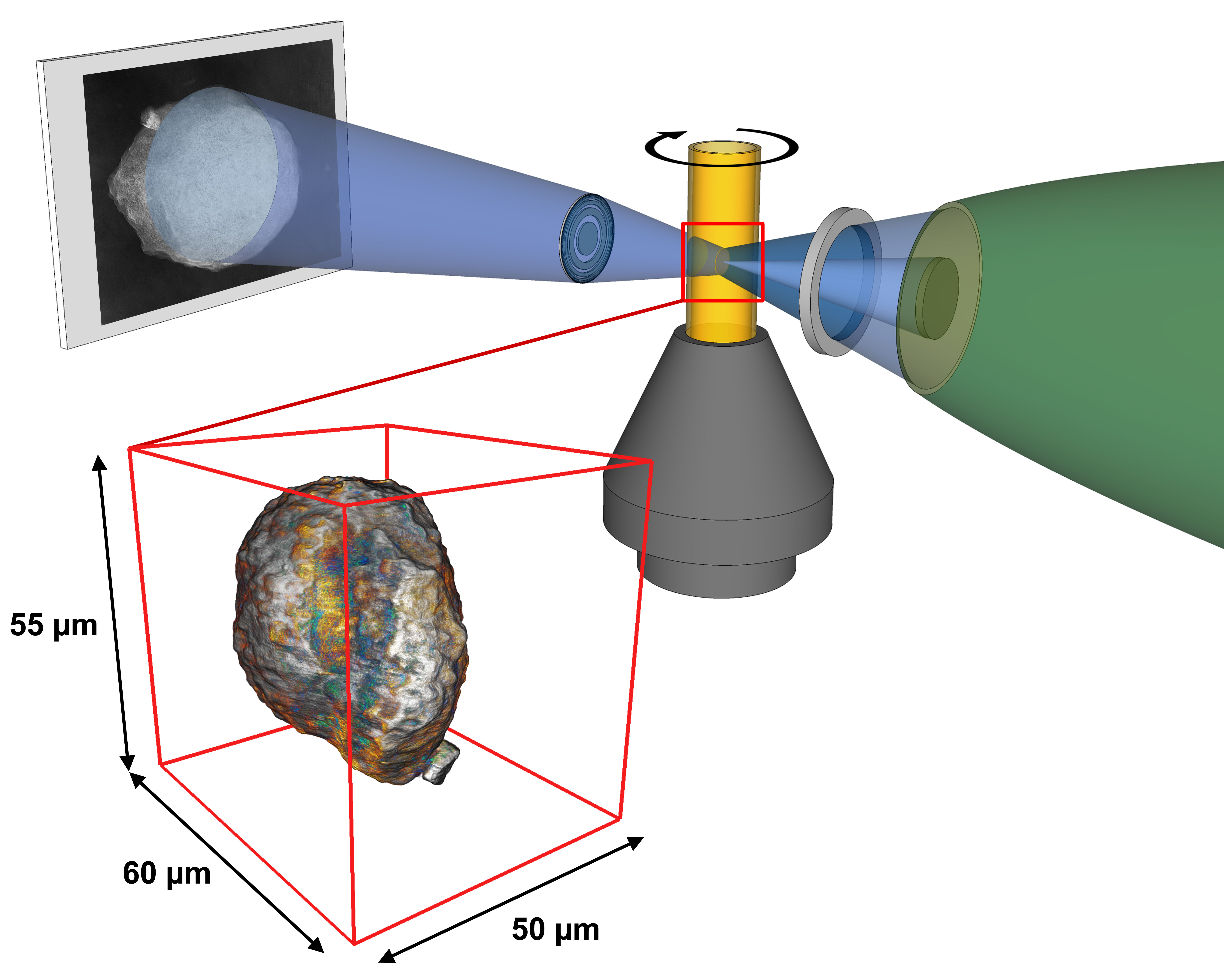
SSRL X-rays are focused to illuminate a small sample of catalysts inside a movable cylindrical holder. A lens magnifies the resulting sample image onto a screen, a CCD camera captures the 2-D image, and software is used to reconstruct a 3-D image of the single catalyst particle from a series of these 2-D images. (Florian Meirer/Utrecht University)
Scientists at SLAC and Utrecht University have identified key problems in the crude oil refining process in an effort to increase the production yield of gasoline.
Their recent experiments at SLAC National Accelerator Laboratory studied catalysts that crack apart the long-chain hydrocarbons in crude oil into smaller, more valuable hydrocarbons like gasoline. The efficiency of this refinement process decreases as the catalysts age.
The researchers used X-ray beams at SLAC’s Stanford Synchrotron Radiation Lightsource to image whole catalyst particles and their internal structure with high resolution – like taking a landscape photograph where you can see a panoramic view and zoom in to see the ants.
To learn more about this research, check out my communications article for SLAC National Accelerator Laboratory.
Morphine is a common narcotic pain reliever with significant side effects (sfxeric/Flickr).
Morphine is a powerful narcotic, commonly used to treat moderate to severe pain from surgery, injury and chronic health conditions like cancer or osteoarthritis. However, morphine has many negative side effects. It can cause drowsiness, nausea, vomiting, dizziness and constipation. More troubling, people can become dependent on it.
According to the Centers for Disease Control and Prevention, narcotic dependence and overdose deaths are a growing health problem in the United States. Narcotic sales quadrupled from 1999 to 2010 and narcotic-related deaths more than tripled from 1999 to 2012. More than 2 million people in the U.S. currently abuse narcotics.
In order to address this problem, researchers have long searched for alternative drugs that effectively relieve pain without inducing dependence as morphine does. A team of researchers now believe they’ve found a good candidate for such a drug, although it will be a long time before this drug, if it proves to be effective, is available in stores.
The LCLS creates the brightest X-ray source on the planet, which researchers need to accurately study the structure of tiny, nanoscale drug compounds.
How Narcotics Work
Narcotics are also called opioid pain relievers, because they block the sensation of pain by binding to opioid receptors within the pain pathway of the brain. A drug can provide pain relief by binding to one of three types of opioid receptors in the brain: delta, mu and kappa. Narcotics like morphine target the opioid mu receptors.
The problem is that long-term use of morphine reduces the number of available opioid mu receptors. As a result, tolerance develops so a higher dose of the drug is needed to achieve the same level of pain relief. Ultimately, prolonged use leads to physical dependence — which is when the neurons adapt to the presence of the drug and function normally only when it’s in the body.
Previous research has shown that administering morphine with another drug that simultaneously blocks the opioid delta receptors prevents this morphine-induced tolerance and dependence. Researchers just need to find the right drug combination with minimal side effects.
Recently, a team of researchers, led by University of Southern California chemistry professor Vadim Cherezov, developed a drug that activates opioid mu receptors while blocking opioid delta receptors. Their drug is derived from a specialized peptide – a naturally occurring chain of amino acids. Their research results were reported in the February issue of Nature Structural and Molecular Biology.
Studying Tiny Crystals
Unfortunately, it is very difficult to study new opioid compounds like the one Cherezov recently developed.
In order to understand the structure of new compounds, scientists usually grow crystals of the compound and then hit them with X-rays. By measuring the angles and intensity of how the X-rays bounce off the crystals, they can produce a three-dimensional picture of the crystal structure.
However, it can be difficult to grow crystals large enough to use this standard method, called X-ray crystallography. Plus, you typically need to freeze the crystal to make it more rigid, rather than study it in natural conditions and temperatures. On top of that, a conventional X-ray beam might blast and destroy the small crystal before you can collect enough data.
Instead, Cherezov’s team used the Linac Coherent Light Source to perform their experiment on tiny crystals at room temperature. LCLS X-ray pulses last just a quadrillionth of a second, or 100 times faster than it takes light to travel the width of a human hair. Yet they are a billion times brighter than a conventional X-ray source.
Using this unique X-ray source with higher-intensity, very short pulses, Cherezov was able to use smaller crystals and still collect terabytes of structural data before the crystals vaporized.
Each crystal the research team prepared was a millionth of a meter and contained many copies of their new opioid compound bound to an opioid receptor. The team placed the crystals in a toothpaste-like gel to simulate the receptors’ natural environment, and then injected a thin stream of this gel into the path of the LCLS X-ray beam.
The resulting structural model was precise enough to show how the new drug molecules bind with the receptor. This atomic-scale map should help scientists develop pain-relieving drugs with fewer negative side effects. It’s likely to to take years, though, before it can be tested in humans.
“This work will provide a solid basis for the design of a new generation of pain relievers with reduced dependency,” Cherezov said in a press release.
Cherezov’s experiment is just one of many performed at the Linac Coherent Light Source.
The LCLS is a Department of Energy User Facility where approximately 600 scientists conduct groundbreaking experiments each year, across many fields, including chemistry, biology, material science, technology and energy science.
Below is a quick post for my former/current high energy physics friends. You can also check out my article for a general audience, which was just published in Symmetry magazine.
The neutrino physics community has wanted to build an accelerator-based, long-baseline neutrino facility for years. But recent efforts appear to be making this exciting experimental program a reality with the formation of the Deep Underground Neutrino Experiment (DUNE) — a truly international collaboration of physicists from 23 countries and 150 institutions.
This worldwide expertise and resources will be needed to make the experiment a reality. DUNE is so challenging that a single nation or continent is unable to do the experiment by itself.
The ambitious experiment will drive a high-intensity, megawatt class neutrino beam from Fermilab through 1300 km of earth to the Sanford Underground Research Facility in South Dakota, where it will be detected by a massive liquid argon time-projection-chamber located deep underground. The plan is to first deploy a 10-kiloton underground detector by 2021, which will later be upgraded to 40-kiloton. A high-resolution detector will also be placed just downstream from the beamline to measure the composition of the neutrino beam as it leaves the Fermilab site.
The principal goal of the experiment is to carry out a comprehensive investigation of neutrino oscillations. Scientists hope to observe CP violation – the asymmetry between matter and antimatter – among neutrinos and compare it to the CP violation seen in quarks and antiquarks. They also aim to determine the ordering of the neutrino masses and to test the three-neutrino paradigm. In addition, extensive neutrino astrophysics and nucleon decay programs are planned using the near and far detectors.
The DUNE collaboration hopes to build this experiment on an aggressive schedule, so you will undoubtedly be hearing more about DUNE soon…
Device used by UC Davis researchers to rapidly concentrate stem cells, which are harvested from surgical irrigation fluid during an orthopedic procedure (Courtesy of SynGen Inc).
About 6 million people in North America suffer bone fractures each year and 5 to 10 percent of these patients are resistant to healing, according to the American Academy of Orthopaedic Surgeons. This means that about half a million Americans annually have fractures that don’t heal. UC Davis researchers are developing an improved surgical therapy for such fractures, using stem cells and innovative technology.
After a broken bone is treated, new bone tissue usually begins to form and connect the broken pieces. However, some bone fractures don’t heal due to a lack of adequate stability, blood flow, or large bone loss. For instance, severe bone fractures that are caused by a high-energy car wreck are more likely not to heal. Several other factors increase the risk of non-healing bones, including older age, diabetes, poor nutrition, use of tobacco, and severe anemia. Traditional treatments to address this problem, such as bone grafts taken from another part of the body, often lead to pain, dysfunctional limbs, and disabilities.
In the last several years, the application of stem cells directly to the wound site has emerged as an improved way to treat non-healing fractures. However, acquiring the necessary stem cells from the patient, a matched donor, or embryo is problematic. Ideally the stem cells come directly from the patient, but this requires a painful surgical procedure with general anesthesia during which a large needle is used to retrieve the stem cells from the hip. In addition, the retrieved stem cells need to be isolated before they can be transplanted back into a patient, so a second surgery is required with a long combined recovery period.
“People come to me after suffering for six months or more with a non-healing bone fracture, often after multiple surgeries, infections and hospitalizations,” said Mark Lee, UC Davis associate professor of orthopaedic surgery, in a press release. “Stem cell therapy for these patients can be miraculous, and it is exciting to explore an important new way to improve on its delivery.”
Mark Lee, UC Davis associate professor of orthopedic surgery (Courtesy of UC Davis).
In their new clinical trial, Lee’s team is testing a new SynGen Inc. device that processes the irrigation fluid obtained during an orthopedic procedure. This irrigation fluid contains abundant mesenchymal stem cells and other factors that can be used to help make new blood vessels and improve wound healing.
During the surgery, the irrigation fluid is aspirated and captured. The fluid is then centrifuged and processed using the new SynGen device, which rapidly isolates a concentration of mesenchymal stem cells in less then 30 minutes. These concentrated stem cells are then delivered to the patient’s fracture during the same surgery. The device is about the size of a food processor, so it can be easily used in an operating room.
“The device’s small size and rapid capabilities allow autologous stem cell transplantation to take place during a single operation in the operating room rather than requiring two procedures over a period of weeks,” said Lee in the press release. “This is a dramatic difference that promises to make a real impact on wound healing and patient recovery.”
The UC Davis researchers are already testing this new surgical treatment on patients. However, it is unclear when this treatment could move into general clinical practice.
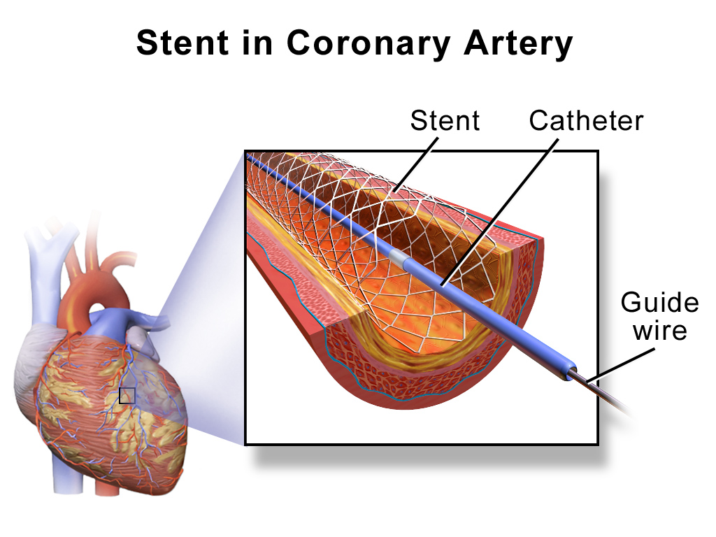
Researchers from Stanford University School of Medicine believe they’ve found a drug for cardiac stents that can more effectively prevent stent complications.
Over a million people in the U.S. each year undergo angioplasty heart surgery using a drug-coated stent to treat blocked arteries, according to the American Heart Association. A stent is a tiny wire mesh tube that is permanently implanted into the artery at the blockage point, creating a scaffold that props open the artery to reduce the chance of a heart attack. However, placement of bare metal stents can themselves damage the artery lining, causing scar tissue to grow and narrow the artery. Known as in-stent stenosis, this typically occurs 3-6 months after the surgical procedure and can lead to chest pain and even heart attacks.
To help prevent in-stent stenosis, doctors use stents coated with drugs that inhibit tissue regrowth to help prevent the blood vessels from reclosing. Unfortunately, these drugs can also inhibit beneficial regrowth of the vessel’s blood lining (endothelium) that aids the healing process. So patients still need to take blood-thinners for up to a year to reduce the risk of a blood clot developing in the stent and blocking the artery. This need for blood thinners is a serious problem for many people with other health issues; for instance, it means they can’t have surgery while taking the medication.
Stanford researchers have now identified a drug to coat cardiac stents that helps prevent in-stent stenosis without affecting the healing of the blood vessel lining. Their new research is described in a paper published this month in the Journal of Clinical Investigation. Dr. Euan Ashley, associate professor of cardiovascular medicine and genetics at Stanford University Medical Center, led the research team.
The researchers first sought to more fully understand the genetic pathways of coronary artery disease using a “big data” computational biology approach. Using data from previous studies, they analyzed large datasets of coronary artery tissue samples and genome information from patients who had developed in-stent stenosis after undergoing angioplasty and stenting. Based on network analyses, the researchers hypothesized that there is an increased risk of in-stent stenosis due to the interplay of two genes, GPX1 and ROS1.
GPX1 deficiency is known to be independently associated with coronary artery disease in humans. However, ROS1 expression is mostly known for its role in highly malignant cancers, such as lung cancers.
“We didn’t know anything about ROS1,” said Ashley in a press release. “It hadn’t been studied in cardiovascular disease. We knew it was an important gene in cancer. We thought, that’s odd, since the growth caused by stents is almost like a tumor.”
They confirmed their theory by performing an extensive series of laboratory experiments using human tissue samples and genetically engineered knockout mice. Some of these studies involved surgically implanting drug-coated stents in mice with clogged arteries. The researchers inhibited the ROS1 genes by coating these stents with crizotinib – a chemotherapy drug used to treat certain ROS1-positive lung cancers. They found that crizotinib inhibited in-stent stenosis without affecting the lining of the blood vessels.
“The major finding of the study is that artery stent disease acts surprisingly like a tumor in the blood vessel wall,” said Ashley in the press release. “Inhibiting it with nonspecific pharmaceutical agents, as we do now, leads to heart attacks from clots caused by lack of endothelial lining on the stent. Whereas, targeting it with the drug we use here, crizotinib, acts much more specifically and inhibits the disease without affecting the endothelium.”
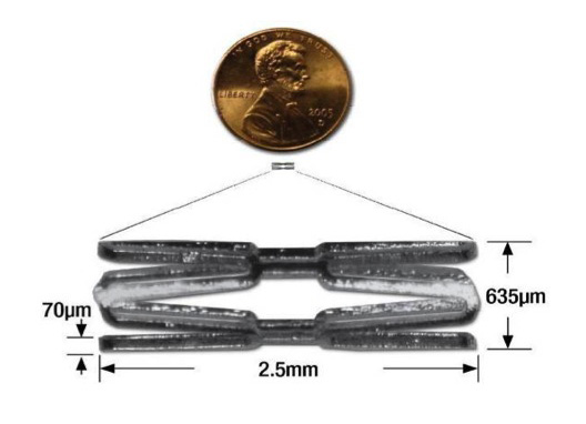
A tiny mouse-sized stent used by Stanford researchers in their mice studies. (Courtesy of Euan Ashley)
Stanford researchers still have a lot more work to do before crizotinib-coated stents will be clinically available. However, this research should translate to the clinic more quickly since crizotinib is already an FDA approved drug.