Millions of people in developing countries lack access to safe drinking water, a primary cause of disease. Now, researchers from Stanford University and SLAC National Accelerator Laboratory have developed a tiny gadget that may help address this huge global health issue.
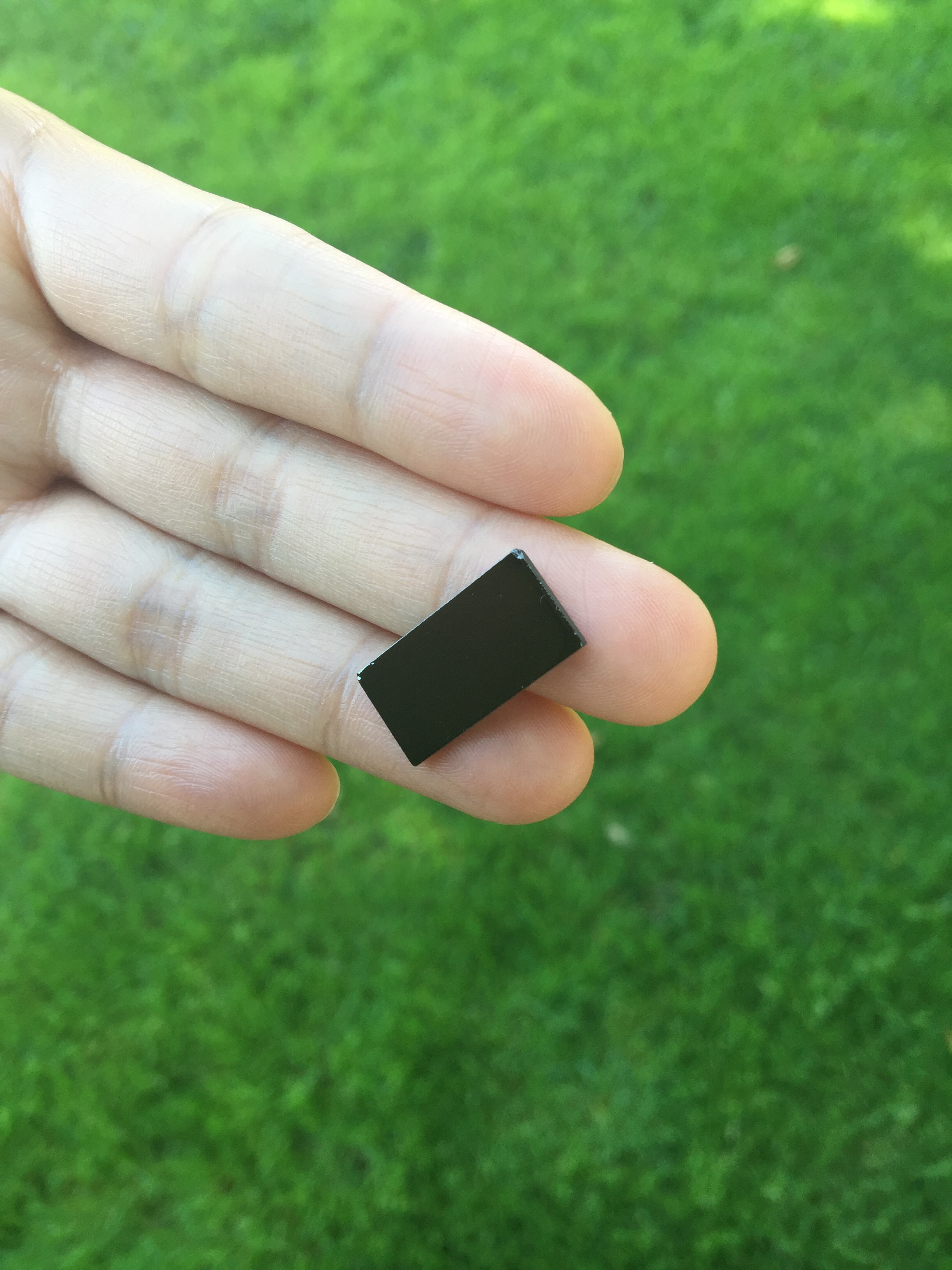
Their device — the size of half a postage stamp — uses solar energy to more efficiently purify water.
Solar energy is already commonly used to disinfect drinking water, particularly in areas with limited fuel to boil water. However, solar disinfection mostly relies on killing pathogens in the water with ultraviolet light, which represents only 4 percent of the sun’s total energy. This slow and inefficient purification process takes six to 48 hours.
The Stanford research team devised a new material that can significantly speed up this process by harvesting the whole spectrum of visible light, which corresponds to over 50 percent of solar energy. As reported this week in Nature Nanotechnology, they were able to disinfect nearly all of the bacteria in a small water sample in just 20 minutes.
“Our device looks like a little rectangle of black glass. We just dropped it into the water and put everything under the sun, and the sun did all the work,” said Chong Liu, PhD, lead author and postdoctoral researcher in materials science and engineering at Stanford, in a recent news release, which describes the device:
Under an electron microscope the surface of the device looks like a fingerprint, with many closely spaced lines. Those lines are very thin films — the researchers call them “nanoflakes” — of molybdenum disulfide that stacked on edge, like the walls of a labyrinth, atop a rectangle of glass.
…
By making their molybdenum disulfide walls in just the right thickness, the scientists got them to absorb the full range of visible sunlight. And by topping each tiny wall with a thin layer of copper, which also acts as a catalyst, they were able to use that sunlight to trigger exactly the reactions they wanted — reactions that produce “reactive oxygen species” like hydrogen peroxide, a commonly used disinfectant, which kill bacteria in the surrounding water.
Although promising, the researchers’ method doesn’t remove chemical pollutants and it has only been tested on three strains of bacteria mixed with less than an ounce of water in the lab. The next step will be to test the device in a real-world stew of contaminants.
This is a reposting of my Scope blog story, courtesy of Stanford School of Medicine.


