Behind the scenes with a Stanford pediatric surgeon

In a new series, “Behind the Scenes,” we’re inviting Stanford Medicine physicians, nurses, researchers and staff to share a glimpse of their day.

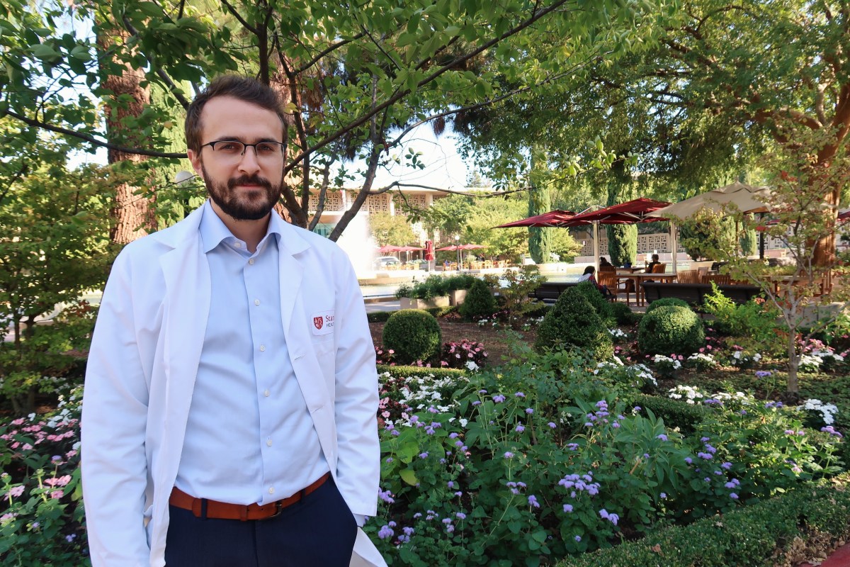
As a science writer, I talk to a lot of health care providers about their work. But I’ve often wondered what it is really like to be a surgeon. So I was excited to speak with pediatric surgeon Stephanie Chao, MD, about her day.

Chao is a pediatric general surgeon, an assistant professor of surgery and the trauma medical director for Stanford Children’s Health. In addition to performing surgeries on children of all ages, she has a range of research interests, including how to reduce gun-related deaths in children and the hospital cost associated with pediatric firearm injuries.

Morning routine
On days that I operate, I get up between 5:50 and 6 a.m., depending on whether I hit the snooze button. I typically don’t eat breakfast. I don’t drink coffee because I don’t want to get a tremor. I’m out the door by 6:30 a.m. and at the hospital by 7 a.m. I usually go by the bedside of the first patient I’m going to operate on to say hi. The patient is in the operating room by 7:30 a.m. and my cases start.

On non-surgical days, it’s more chaotic. I have a 3-year-old and 1-year-old. So every day there’s a jigsaw puzzle as to whether my husband or I stay to get the kids ready for preschool, and who comes home early.

Part of Stephanie Chao’s day involves checking on patients, including this newborn.

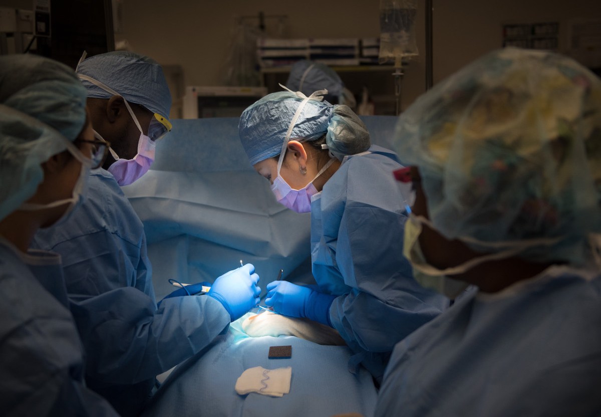
In the operating room
The operating room is the place where I have the privilege of helping children feel better. It’s a very calming place, like a temple. When I walk through the operating room doors, the rest of the world becomes quiet. Even if it is a high-intensity case when the patient is very sick, I know there is a team of nurses, scrub techs and anesthesiologists used to working together in a well-orchestrated fashion. So even when the unexpected arises, we can focus on the patient with full confidence that we’ll find a solution.

There are occasions when babies are so sick that we need silence in the operating room. Everyone becomes hyper-attuned to all the beeps on the monitors. When patients are not as critically sick, I often play a Pandora station that I created called “Happy.” I started it with Pharrell Williams’ “Happy” and then Pandora pulled in other upbeat songs, including a bunch of Taylor Swift songs, so everyone thinks I’m a big Taylor Swift fan.

The OR staff call me by my first name. I believe that if everyone is relaxed and feels like they have an equal say in the procedure, we work better as a well-oiled machine for the benefit of the patient.

“The OR staff call me by my first name,” Stephanie Chao said.

Favorite task
Some of the most rewarding times of my day are when I sit down with patients and their families to hear their concerns, to reassure them and to help them understand what to expect — and hopefully to make a scary situation a little less so. As a parent, I realize just how hard it is to entrust one’s child completely in the hands of another. I also like to see patients in the hospital as they’re recovering.

Favorite time
The best part of the day is when I come home. When I open the door into the house, my kids recognize that sound and I hear their little footsteps as they run towards the door, shrieking with joy.

Evening ritual
When my husband and I get home, on nights I am not on call, I cook dinner in the middle of the chaos of hearing about the kids’ day. Hopefully, we “sit down” to eat by 6:20 or 6:30 p.m., and I mean that term loosely. It’s a circus, but eventually everyone is somewhat fed.

And then we do bath time and bedtime. There’s a daily negotiation with my three-year-old on how many books we read before bed. On school nights, she’s allowed three books but she tries to negotiate for 10.

Eventually, we get both kids down for the night. Then my husband and I clean up the mess of the day and try to have a coherent conversation with each other. But by then both of us are exhausted. I try to log on again to finish some work, read or review papers. I usually go to sleep around 11 p.m.

Managing it all
When I can carve out time to do relaxing things for myself, like go to the gym, that is great. But it’s rare and I remind myself that I am blessed with a job that I love and a wonderfully active family.

The result sometimes feels like chaos, but I don’t want to wish my life away waiting for my kids to get older and for life to get easier. Trying to live in the moment, and embracing it, is how I find balance.

Photos by Rachel Baker

This is a reposting of my Scope blog story, courtesy of Stanford School of Medicine.

“Poor air quality affects everyone” — How to protect yourself and clean the air

I remember when you could ride BART for free on a “Spare the Air” day, when smog was expected to reach unhealthy levels based on standards set by the Environmental Protection Agency. Now, there are too many of these days — 26 in the Bay Area last year — to enjoy that perk.

This bad air is making us sick, according to Stanford allergy specialist and clinical associate professor Sharon Chinthrajah, MD. In a recent episode of the Sirius radio show “The Future of Everything,” she spoke with Stanford professor and radio host Russ Altman, MD, PhD, about how we can combat the negative health impacts of air pollution.

“Poor air quality affects everybody: healthy people and people with chronic heart and lung conditions,” said Chinthrajah. “And you know, in my lung clinic I see people coming in with exacerbations of their underlying lung diseases like asthma or COPD.”

On Spare the Air days, Chinthrajah said even healthy people can suffer from eye, nose, throat and skin irritations caused by air pollution. And the health impacts can be far more serious for her patients. So she tells them to prepare for bad air quality days and to monitor the air quality index (AQI) in their area, she said.

The AQI measures the levels of ozone and other tiny pollutants in the air. The air is considered unhealthy when the AQI is above 100 for sensitive groups — like people with chronic illnesses, older adults and children. It’s unhealthy for everyone when the AQI is above 150.

On these unhealthy air days, Chinthrajah recommends taking precautions:

  • Limit the time you spend outdoors.
  • When outside, use a well-fitted air mask that filters out pollutants larger than 2.5 microns (which is about 20 times smaller than the thickness of an average human hair).
  • When driving, recirculate the air in your car and keep your windows closed.
  • Stay hydrated.
  • Once inside, change your clothes and take a quick shower before you go to bed, removing any air particulates that collected on you during the day.

In the radio show, Chinthrajah explained that published studies by the World Health organization and others demonstrate that people who live in developing countries like India and Asia — where they suffer poor air quality many days of the year — have a shortened life span.

“You know, there’s premature deaths. There’s exacerbation of underlying lung issues and cardiovascular issues. There’s more deaths from heart attacks and strokes in countries where there is poor air quality,” she said.

She admitted that it is difficult to definitively say these health problems are due to poor air quality — given the other problems in the developing country

es like limited access to clean water, food and health care — but she thinks poor air quality is a major contributor.

Chinthrajah said she believes we need to address the problem of air pollution at a societal level. And that means we need to target cars that burn fossil fuel, which account for much of the air pollution in California, she said. Instead, we need to move towards using public transportation and electric vehicles, as well as generating electricity from clean energy sources like solar, wind and water.

She noted that California is now offering a $9,5000 subsidy to qualifying low-income families to purchase low emission vehicles like all-electric cars or plug-in hybrids, on top of the standard federal and state rebates.

“So it seems like an overwhelming, daunting task, right? But I think we each have to take ownership of what we can do to reduce our carbon footprint. And then lobby within our local organizations to create practices that are sustainable,” she said.

Chinthrajah hopes that addressing air pollution and energy consumption at a societal level will lead to less asthma and other health problems, she said.

Image by U.S. Environmental Protection Agency 

This is a reposting of my Scope blog story, courtesy of Stanford School of Medicine.

Designing buildings to improve health

Are the buildings that we live and work in stressing us out?

The answer is probably yes, according to Stanford engineer Sarah Billington, PhD, and her colleagues. They also believe this stress is taking a significant toll on our mental and physical health because Americans typically spend almost 90% of their lives indoors.

During a recent talk at a Stanford Reunion Homecoming alumni celebration, Billington described a typical noisy office cut off from nature and filled with artificial light and artificial materials. This built environment makes workers feel stress, anxiety and distraction, which reduces their productivity and their ability to collaborate with others, she explained.

Now, Billington’s multidisciplinary research team is working to design buildings that instead reduce stress and increase a sense of belonging, physical activity and creativity.

Their first step is to measure how building features — such as airflow, lighting and views of nature — affect human well-being. They are quantifying well-being by measuring levels of stress, belonging, creativity, physical activity and environmental behavior.

In a preliminary online study, the team showed about 300 participants pictures of different office environments and asked them to envision working there at a new job. Across the board, the fictitious work environment was viewed as important to well-being.

“In eight out of the nine things that we were looking at, there were statistically significant increases in their sense of belonging, their self-efficacy and their environmental efficacy when they believed they were going to be working in an environment that had natural materials, natural light or diverse representations,” said Billington.

The researchers are now expanding this work by performing larger lab studies and designing future field studies. They plan to collect data from “smart buildings,” which use high-tech sensors to control the heating, air conditioning, ventilation, lighting, security and other systems. The team also plans to collect data from personal devices such as smartwatches, smartphones and laptops.

By analyzing all of this data, they plan to infer the participants’ behaviors, emotions and physiological states. For example, the researchers will use the building’s occupancy sensors to detect if a worker is interacting with other people who are nearby. Or they will figure out someone’s stress level based on how he or she uses a laptop trackpad and mouse, Billington said.

Stanford computer scientist Pablo Paredes, PhD, who collaborates on the project, explained in a paper how their simple model of arm-hand dynamics can detect stress from mouse motion. Basically, your muscles get tense and stiff when you’re stressed, which changes how you move a computer mouse.

Next, the team plans to use statistical modeling and machine learning to connect these human states to specific building features. They believe this will allow them to design better buildings that improve the occupants’ health.

The researchers said they intend to bring nature indoors by engineering living walls with adaptable acoustic and thermal properties.

They also plan to incorporate dynamic digital displays — such as a large art display on the wall or a small one on an individual’s personal devices — that reflect occupant activity and well-being. For example, a digital image of a flower might represent the energy level of a working group based on how open the petals are, and this could nudge their behavior, Billington said in the talk.

“Our idea is, what if we could make our buildings shape us in a positive way and keep improving over time?” Billington said.

Photo by Nastuh Abootalebi

This is a reposting of my Scope blog story, courtesy of Stanford School of Medicine.

Eponym debate: The case for biologically-descriptive names

Naming a disease after the scientist who discovered it, like Hashimoto’s thyroiditis or Diamond-Blackfan anemia, just doesn’t work anymore, some physicians say.

A main argument against eponyms is that plain-language names — which describe the disease symptoms or underlying biological mechanisms —  are more helpful for patients and medical trainees. For example, you can probably out a bit about acquired immunodeficiency syndrome (AIDS), whooping cough or pink eye just from their names.

“The more obscure and opaque the name — whether due to our profession’s Greek and Latin fetish or our predecessors’ narcissism — the more we separate ourselves from our patients,” says Caitlin Contag, MD, a resident physician at Stanford.

Stanford endocrinologist Danit Ariel, MD, agrees that patients are often confused by eponyms.

“I see this weekly in the clinic with autoimmune thyroid disease. Patients are often confusing Graves’ disease with Hashimoto’s thyroiditis because the names mean nothing to them,” says Ariel. “So when I’m educating them about their diagnosis, I try to use the simplest of terms so they understand what is going on with their body.”

Ariel says she explains to her patients that the thyroid is overactive in Graves’ disease and underactive in Hashimoto’s.

Ariel says she believes using biological names also helps medical students better understand the underlying mechanisms of diseases, whereas using eponyms relies on rote memorization that can hinder learning. “When using biologically-descriptive terms, it makes inherent sense and students are able to build on the concepts and embed the information more effectively,” Ariel says.

Medical eponyms are particularly confusing when more than one disease is named after the same person, Contag argues. For example, neurosurgeon Harvey Williams Cushing, MD, has 12 listings in the medical eponym dictionary. 

Stanford resident physician Angela Primbas, MD, agrees that having multiple syndromes named after the same person is confusing. She says it’s also confusing to have diseases named differently in different countries. In fact, the World Health Organization has tried to address this, along with other issues, by providing best-practice guidelines for naming infectious diseases. (Genetic disorders, however, lack a standard convention for naming.)

In addition, Primbas said she thinks naming a disease after a single person is an oversimplification of a complex story. “Often many people contribute to the discovery of a disease process or clinical finding, and naming it after one person is unfair to the other people who contributed,” she says. “Plus, it’s often disputed who first discovered a disease.”

Also, few disease names recognize the contributions (or suffering) of women and non-Europeans. And some eponyms are decidedly problematic, like those named after Nazi doctors. A famous example is Reiter’s syndrome named for Hans Reiter, MD, who was convicted of war crimes for his medical experiments performed at a concentration camp.

“Reiter’s syndrome is now called reactive arthritis for the simple reason that Reiter committed atrocities on other human beings to conduct his ‘science.’ Such people should not have their name tied to a profession that espouses the principles of beneficence and nonmaleficence,” says Vishesh Khanna, MD, a resident physician at Stanford. He says medicine is swinging away from using these controversial eponyms to describe them on the basis of their biology instead.

Personally, Khanna also admits that naming a disease after himself wouldn’t sit well.

“Receiving credit for discovering something can certainly be a wonderful feather in a physician’s career cap, but the thought of actually naming a disease after myself makes me cringe,” says Khanna. “Patients and doctors would utter my name every time they had to bring up a disease.”

Such sentiments may be why Contag’s example of a good disease name — cyclic vomiting syndrome — is in plain English. Was no one eager to lend his or her name to it?

While the debate over medical eponyms continues, Khanna suggests a potential solution. “Perhaps a reasonable approach to naming going forward is to allow the use of already established eponyms without dubious histories, while only naming newly discovered diseases based on pathophysiology,” he says.

Everyone I spoke with agrees that changing the medical eponyms will only happen slowly, if at all, since it is difficult to change language. However, it can be done, according to Dina Wang-Kraus, MD, a Stanford resident in psychiatry and behavioral sciences.

“I looked through our diagnostic manual and we do not have diseases named after people in psychiatry. This shift happened quite some time ago so as to avoid confusion and to allow clinicians from all over the world to have a unified language,” says Wang-Kraus. “In psych, we often say that we wish other specialties would adopt a universal nomenclature too.”

This is the conclusion of a series on naming diseases. The first part is available here.

Photo by 4772818

This is a reposting of my Scope blog story, courtesy of Stanford School of Medicine.

Eponym debate: The case for naming diseases after people

Is it better to name a genetic disorder Potocki-Lupski syndrome or the 17p11.2 duplication syndrome? What about Addison’s disease as opposed to adrenal insufficiency? Or Tay-Sachs disease versus hexosaminidase alpha-subunit deficiency (variant B)?

If you have a strong opinion about which is preferable, you aren’t alone: there is an ongoing controversy on how to name diseases. In Western science and medicine, a long-standing tradition is to name a disease after a person. However, many physicians now argue that these eponyms should be abandoned for biologically-descriptive names.

First, a bit about how eponyms are created.

Although the media sometimes comes up with a catchy name that sticks, like swine flu, diseases are typically named by scientists when they first report them in scientific publications.

Oftentimes, diseases are named after prominent scientists who played a major role in identifying the disease. The example that leaps to my mind is Hodgkin’s disease — a type of cancer associated with enlarged lymph nodes — because I was diagnosed and treated for Hodgkin’s at Stanford years ago. Hodgkin’s disease was named after Thomas Hodgkin, an English physician and pathologist who described the disease in a paper in 1832.

Less frequently, diseases are named after a famous patient. For example, amyotrophic lateral sclerosis (ALS), commonly known as Lou Gehrig’s disease, was named after the famous New York Yankee baseball player who was forced to retire after developing the disease in 1939.

As these examples show, one of the reasons to keep eponyms is that they are embedded with medical traditions and history. They include some kind of story. And, oftentimes, they honor key people associated with the disease.

“I think the people who discover these conditions deserve recognition,” explains Angela Primbas, MD, a resident physician at Stanford. “I don’t think the medical community would know their names otherwise.”

Some physicians also feel eponyms bring color to medicine. “The use of eponyms in medicine, as in other areas, is often random, inconsistent, idiosyncratic, confused, and heavily influenced by local geography and culture. That is part of their beauty,” writes Australian medical researcher Judith Whitworth, MD, in an editorial in BMJ.

Other proponents of eponyms are more practical. They argue that eponymous disease names provide a convenient shorthand for doctors and patients.

Medical eponyms are also widely used by patients, physicians, textbooks and websites. According to a dictionary of medical eponyms, thousands of eponyms are used throughout the world particularly in the United States and Europe. They are even prominent in the World Health Organization’s international classification of diseases.

So is a massive effort to purge these eponyms worth it, or even realistic?

“There are certainly examples where eponymous disease names are so inculcated in medical vernacular that changing them to a pathology-based name might not be worth the effort,” says Vishesh Khanna, MD, a resident physician at Stanford. He gives the examples of Alzheimer’s disease and Crohn’s disease.

Jimmy Zheng, a medical student at Stanford, agrees that eponyms are here to stay. “At the level of medical school, eponyms are broadly dispensed in class, in USMLE study resources and in our clinical training,” Zheng says. “While some clinicians have called for the complete erasure of eponyms, this is unlikely to happen.”

Zheng and Stanford neurologist Carl Gold, MD, recently assessed the historical trends of medical eponym use in neurology literature. They also surveyed neurology residents on their knowledge and attitude towards eponyms. Their study’s findings were published in Neurology.

“Regardless of ‘should,’ our analyses demonstrate that eponyms are increasingly prevalent in the scientific literature and that new eponyms like the Potocki-Lupski syndrome continue to be coined,” Gold says. “Despite awareness of both the pros and cons of eponyms, the majority of Stanford neurology trainees in our study reported that historical precedent, pervasiveness and ease of use would drive the continued use of eponyms in neurology.”

So the debate rages on. According to my informal and small survey, some Stanford physicians favor eliminating eponymous disease names — stay tuned to find out why.

This is the beginning of a two-part series on naming diseases. The conclusion will appear this week.

Photo via Good Free Photos

This is a reposting of my Scope blog story, courtesy of Stanford School of Medicine.

Measuring depression with wearables

Depression and emotional disorders can occur at any time of year — and do for millions of Americans. But feeling sad, lonely, anxious and depressed may seem particularly isolating during this holiday season, which is supposed to be a time of joy and celebration.

A team of Stanford researchers believes that one way to work towards ameliorating this suffering is to develop a better way to quantitatively measure stress, anxiety and depression.

“One of the biggest barriers for psychiatry in the field that I work in is that we don’t have objective tests. So the way that we assess mental health conditions and risks for them is by interview and asking you how do you feel,” said Leanne Williams, MD, a professor in psychiatry and behavioral sciences at Stanford, when she spoke at a Stanford Reunion Homecoming alumni celebration.

She added, “Imagine if you were diagnosing and treating diabetes without tests, without sensors. It’s really impossible to imagine, yet that is what we’re doing for mental health, right now.”

Instead, Stanford researchers want to collect and analyze data from wearable devices to quantitatively characterize mental states. The multidisciplinary team includes scientists from the departments of psychiatry, chemical engineering, bioengineering, computer science and global health.

Their first step was to use functional magnetic resonance imaging to map the brain activity of healthy controls compared to people with major depressive disorder who were imaged before and after they were treated with antidepressants.

The researchers identified six “biotypes” of depression, representing different ways brain circuitry can be disrupted to cause specific symptoms. They classified the biotypes as rumination, anxious avoidance, threat dysregulation, anhedonia, cognitive dyscontrol and inattention.

“For example, threat dysregulation is when the brain stays in alarm mode after acute stress and you feel heart racing, palpitations, sometimes panic attacks,” presented Williams, “and that’s the brain not switching off from that mode,” Williams said.

The team, which includes chemical engineer Zhenan Bao, PhD, then identified links between these different brain biotypes and various physiological differences, including changes in heart rate, skin conductance, electrolyte levels and hormone production. In particular, they found correlations between the biotypes and production of cortisol, a hormone strongly related to stress level.

Now, they are developing a wearable device — called MENTAID — that measures the physiological parameters continuously. Their current prototype can already measure cortisol levels in sweat in agreement with standard laboratory measurements. This was an incredibly challenging task due to the extremely low concentration and tiny molecular size of cortisol.

Going forward, they plan to validate their wearable device with clinical trials, including studies to assess its design and user interface. Ultimately, the researchers hope MENTAID will help prevent and treat mental illness — for example, by better predicting and evaluating patient response to specific anti-depressants.

Photo by Sora Sagano

This is a reposting of my Scope blog story, courtesy of Stanford School of Medicine.

X-rays shed light on how anti-asthmatic drugs work

A new study uncovers how a critical protein binds to drugs used to treat asthma and other inflammatory diseases.

By studying the crystal structure of an important protein when it was bound to two drugs widely prescribed to treat asthma, an international team of scientists has discovered unique binding and signaling mechanisms that could lead to the development of more effective treatments for asthma and other inflammatory diseases.

The protein, called cysteinyl leukotriene receptor type 1 (CysLT1R), controls the dilation and inflammation of bronchial tubes in the lungs. It is therefore one of the primary targets for anti-asthma drugs, including the two drugs studied: zafirlukast, which acts on inflammatory cells in the lungs, and pranlukast, which reduces bronchospasms due to allergic reactions.

Using the Linac Coherent Light Source (LCLS) X-ray free-electron laser at the Department of Energy’s SLAC National Accelerator Laboratory, the team bombarded tiny crystals of CysLT1R-zafirlukast with X-ray pulses and measured its structure. They also used X-rays from the European Synchrotron Radiation Facility in Grenoble, France to collect data about CysLT1R-pran crystals. They published their findings in October in Science Advances.

The researchers gained a new understanding of how CysLT1R interacts with these anti-asthma drugs, observing surprising structural features and a new activation mechanism. For example, the study revealed major differences between how the two drugs attached to the binding site of the protein. In comparison to pranlukast, the zafirlukast molecule jammed open the entrance gate of CysLT1R’s binding site into a much wider configuration. This improved understanding of the protein suggests a new rationale for designing more effective anti-asthma drugs.

The study was performed by a collaboration of researchers at SLAC; Moscow Institute of Physics and Technology, Russia; University de Sherbrooke, Canada; University of Southern California; Research Center Juelich, Germany; Universite Grenoble Alpes-CEA-CNRS, France; Czech Academy of Sciences, Czech Republic; and Arizona State University.

Citation: Aleksandra Luginina et al., Science Advances, 09 October 2019 (10.1126/sciadv.aax2518).

For questions or comments, contact the SLAC Office of Communications at communications@slac.stanford.edu.

Image caption: Using X-rays, researchers uncovered details about two drugs widely prescribed to treat asthma: pranlukast (shown up top) and zafirlukast (shown beneath). Their results revealed major differences between how the two drugs attached to the binding site of the receptor protein. In comparison to pranlukast, the zafirlukast molecule jammed open the entrance gate of protein’s binding site into a much wider configuration. (10.1126/sciadv.aax2518)

This is a reposting of my SLAC news story, courtesy of SLAC Linear Accelerator Laboratory.

Testing infants’ blood may predict psychological health, study finds

Many of us know that a lipid panel — a simple blood test that measures the levels of cholesterol and fats in the blood — can help predict the risk of heart disease in adults.

What may be more surprising is a Stanford study has now shown that the levels of cholesterol and fat in an infant’s blood can predict that child’s social and emotional development, as recently reported in Psychological Science.

The researchers analyzed data compiled by the Born in Bradford project, which followed children born in the United Kingdom city of Bradford between March 2007 and December 2010.

The Stanford team examined the levels of high-density lipoproteins (HDL) known as “good cholesterol,” very-low-density lipoproteins (VLDL) known as “bad cholesterol” and triglycerides in the umbilical cord blood of 1,369 newborns. Unlike the placenta, all the cells in cord blood are from the fetus.

They then correlated the blood results with the children’s psychological status — including their self-awareness, emotional regulation and interpersonal relationships — as measured five years later by their teachers using standard tests.

The study showed children born with higher levels of HDL, lower levels of VLDL and lower levels of triglycerides were more likely to receive higher teacher ratings than their peers with lower “good cholesterol.”

“It is surprising that from early in life, these easily accessible and commonly examined markers of blood lipid levels have this predictive correlation for future psychological outcomes,” said Erika Manczak, PhD, in a recent Stanford news release. “What our study showed is really an optimistic finding because lipids are relatively easy to manipulate and influence.” Manczak participated in the research as a postdoctoral fellow in psychology at Stanford and is now an assistant professor of psychology at Denver University.

The study, so far, has demonstrated only correlations, not causations. But the findings were consistent across different ethnic and socioeconomic backgrounds and both sexes, where the study participants were 38% white British, 51% Pakistani British, 11% of other ethnicity and 52% male. The associations also held regardless of the mother’s psychological or physical health during pregnancy or the children’s physical health, body mass or neurodevelopmental status.

“The fact that the only solid predictor for the Born in Bradford children’s psychosocial competency assessment scores was their fetal lipid levels really argues in favor of a connection between the two,” Manczak said in the release. “Now we need to find out what exactly this connection may be.”

In the paper, the authors suggest some potential explanations, noting that lipids are involved in many biological processes important to psychological health, such as brain development and inflammation. If future work confirms their findings, they hope lipid screening can help identify and guide treatment for children who are prone to mental illnesses.

Photo by ThorstenF

This is a reposting of my Scope blog story, courtesy of Stanford School of Medicine.

Publish or perish: The cost of reformatting academic papers

You’ve probably heard the expression “publish or perish,” which describes the pressure to publish research in order to succeed in an academic career.

You’d think that conducting the research needed to write a paper would be the hard part — and it is. But publishing isn’t easy either, a new Stanford-led study in PLOS One emphasizes. Even top researchers often have to submit papers to multiple journals before getting one to accept it. This process is very time consuming and frankly a bit painful for most authors.

The new study quantifies the pain and cost of a key part of this resubmission process — reformatting the manuscript to another journal’s guidelines.

“All researchers have wasted an inordinate amount of time reformatting papers to another journal’s specific requirements for things like word count, font and figure limits, which is entirely separate from improving the scientific content,” said Sidhartha Sinha, MD, a gastroenterologist and researcher at Stanford. “As medical researchers, we should be spending this time on actual research and patient care, not on adhering to seemingly arbitrary and highly variable formatting requirements.”

So just how detailed are these formatting guidelines? Sinha shared one of his favorite absurd examples taken from a top medical journal: “Type decimal points midline (ie, 23·4, not 23.4). To create a midline decimal on a PC: hold down ALT key and type 0183 on the number pad, or on a Mac: ALT shift 9.”  

He suggests that these rules shouldn’t matter during the initial submission and review process, particularly given that the rejection rate for biomedical journals is 62% on average and over 90% at top tier journals.

Sinha and his colleagues were inspired to study this problem after years of feeling frustrated with the current inefficient process. Although everyone complains about it, very little actual research has been done on the topic, he said.

The team of physicians and editors randomly selected 96 journals focused on basic and clinical biomedical research. They then randomly selected three recently published, original research articles from each journal and sent their survey to the first or corresponding author. A total of 203 authors filled out the survey.

“We had a very high response rate of 72%, which shows that we struck a chord with researchers because it is such a huge problem,” said Sinha. “In fact, only 12% of authors indicated satisfaction with the current resubmission process.”

The survey asked about the time spent by the participating authors and their entire research team to reformat resubmissions for their recent paper. Participants also gave input on the overall reformatting process and how it could be improved.

The study found that most of the 203 authors spent 1 to 3 days or more on reformatting alone, which delayed resubmissions by over two weeks in most instances and up to three months for 20% of the manuscripts.

“It’s not that they are spending three months on reformatting, but they get sidetracked with grant deadlines or other research pursuits,” explained Sinha. “In fact, I currently have one manuscript that is indefinitely on the back burner because I’ve already submitted it a few times and have other research priorities .”

Based on their survey results, the authors estimated that the total time spent reformatting the 2.3 million scientific articles published annually translates into a global cost of over $1 billion. And Sinha said the actual cost is likely much higher — since they assumed, for example, a first-year postdoc salary of $48,000 for all authors even though senior authors make significantly more — and much of this cost is funded by taxpayers’ dollars.

In the paper, the authors make some recommendations — including adopting a universal format-free initial submission policy. However, they primarily hope their study will start a discussion about how to improve the existing broken process, Sinha said.

“There are trends towards minimizing formatting requirements, but there is still much room for improvement,” said Sinha. “I’d like editors from each field to get together and agree on generalized formatting guidelines. For example, maybe brief reports are 3,000 words and original research articles are 6,000 words. And it might be different for radiology and cell biology journals. But we can find a better way to disseminate research faster and more cost-effectively.”

So, like me, are you wondering how much time his team spent on reformatting this paper on publication inefficiencies?  “We kept track and we spent just over 25 hours on reformatting before it was accepted. We hope this paper helps change this in the future,” Sinha said.

Photo by Nic McPhee

This is a reposting of my Scope blog story, courtesy of Stanford School of Medicine.

A twisty career path to improve care for smokers

When Jason Melehani, MD, PhD, grew up in a small town in the Sierra Nevada foothills, he didn’t know any scientists or doctors.

That all changed when he went to college at the University of California, Los Angeles and discovered the world of research. Now, Melehani is a resident in internal medicine at Stanford and his career path, twisty though it may seem, is headed to a future of helping people who have struggled with tobacco use.

At UCLA as a freshman, he joined a lab and began investigating an unusual parasite called Trypanosoma brucei that is transmitted via the bite of a tsetse fly to humans and cattle, causing an often fatal sleeping sickness in Saharan Africa.

“African sleeping sickness exclusively affects very impoverished regions of the world, so there wasn’t much interest from the pharmaceutical industry to develop medicines for this disease,” said Melehani. “A new therapy was recently approved, but it was spearheaded by a nonprofit initiative.”

This research experience inspired his career plan — with the ultimate goal of developing therapies for diseases affecting socioeconomically disadvantaged populations.

First, Melehani headed to the University of North Carolina, Chapel Hill, to earn both a medical degree and doctorate in pharmacology. This program included two years of preclinical medical courses, four years of research and two years of clinical training.  

After completing the MD-PhD program, Melehani took an unconventional approach.  

“Developing new treatments for patients is incredibly challenging especially from an academic lab. You can get things started, but there is a whole world of skills and people required to take things all the way to the clinic,” said Melehani. “I felt like I was experiencing only a thin sliver of the entire process in a research lab.”

To broaden his exposure, he next worked as a fellow at a venture capital firm in North Carolina focusing on healthcare and biotechnology.

“In seven months, I evaluated 500 companies and helped pick the most promising ones, which each received an investment of between half a million to eight million dollars,” said Melehani. “I worked with leaders of major healthcare organizations who valued my opinion despite my junior position. I learned a lot about how new drugs are developed and the role venture capital plays.”

The contacts and insights he gained through this venture capital training and a separate internship in the pharmaceutical industry will likely come in handy in the future when he is running his own academic research lab. “My hope is that this training will help me better select and position future discoveries so I can move them out of my lab to startup companies and ultimately to patients,” he said.

Even at Stanford, Melehani is making his own path. Melehani has applied to do fellowship training next year in both rheumatology and pulmonary medicine, which no one has done before in recent memory.

Melehani plans to research how smoking tobacco affects the immune system and leads to severe health consequences, such as chronic obstructive pulmonary disease, rheumatoid arthritis or heart disease.

“Smoking has disastrous immediate and long term effects on nearly every system in the body,” he said. “And it’s deeply tragic because 90% of people who smoke start before the age of 18 and it’s highly addictive. So even though 70% of people want to quit, the success rates are dismal — around 10%.”

The health impacts of smoking have been on Melehani’s mind for a long time. Many of his friends started smoking in high school. He was exposed to a lot of patients in North Carolina who were smokers. And now at Stanford, he sees many patients who are former smokers and dealing with a wide range of health problems.

Smoking fits his goal of addressing a major socioeconomic health problem— the highest rates of smoking in the United States are in the poorest areas with the lowest education rates. And these are the people who don’t have the resources to face the disastrous health consequences that result, he said.

Melehani hopes to tackle this problem by running his own lab at Stanford, doing fundamental research into how the immune system is affected by cigarette smoke and turning that research into meaningful changes in medical care for his patients.

For now, he is focusing on his patients and getting through his night shifts in the intensive care unit.

Photo of Jason Melehani by Margarita Gallardo

This is a reposting of my Scope blog story, courtesy of Stanford School of Medicine